Category Archives: Treatment

New Concerns Have Sprung

image

Chance greeting me as I drive up

The days are finally feeling like spring!  The grass is green, the blankets put away, the sun is shining, and the horses are shedding.

Chance is continuing to gain weight, although as I said in the previous post, he still needs to put on a good 50-75 lbs.  As the days continue to get warmer, Chance’s arthritis seems to become more manageable for him; his stride is longer and he runs around (mostly after Lucky) more frequently.

Unfortunately, when the farrier came out about two weeks ago Chance was too stiff to get his back right shoe on. The farrier decided to come back out to try and re-shoe him and, during that time in between, Chance must have tweaked it…AGAIN! 

 While Chance did not have a shoe on his back right I kept it wrapped to provide some protection and also even out all of his hooves.  However, when I arrived I noticed that Chance was significantly twisting his back right leg inward at the walk & it had some swelling.  The swelling was not horrible but it was noticeable. I cold hosed his leg for about 45 minutes while I groomed him & gave him a dose of Equinox (pain medication) and Ulcer Guard.  I put on his back leg wrap to help with reducing the swelling and provide some extra support.  Chance did his neck stretches effortlessly and was baring weight on his back hind. 

But as I was grooming him I noticed, on the left side his chest, he had patches of hair loss and dandruff.  The area did not look red or inflamed, nor did it seem itchy or painful.  So I continued grooming him and decided to put a call into the vet to come and check his leg and the hair loss.

Of course, I turned to Google to try and find out what exactly could be the cause of the patches of hair loss.

image

According to a handful of sources, there are a few possibilities for hair loss- mites or Lice, a vitamin deficiency, rain rot or crud, or even just his natural shedding tendency. A skin scrape would help to confirm what may be the cause. 

 As for the swelling of Chance’s back right leg, I decided to call our previous vet who collected and injected Chance’s DDFT with stem cells to heal the hole in his tendon. We have some stem cells left over and I wanted to see if injecting his leg again would be of any benefit.  I also would like to get an ultrasound recheck to ensure that there is not another injury to his DDFT tendon sheath again.

The twisting of his back hind leg is worrisome as well.  

Everything I have read about EPM states that horses can have a relapse in symptoms after treatment is complete. My concern is that the twisting are due to the neurological symptoms coming back since Chance’s EPM treatment has been finished for a little over two weeks…. 

 Our current vet believes that Chance’s ataxia and twisting is not due to EPM but his cervical spine instead.  Could the twisting be worse due to the swelling of his hind leg?  Or is the swelling and the twisting two separate issues all together?

It Just Keeps Getting Better & Better

Two days ago Chance’s vet came out to do a follow up and to give him and Luck their Spring shots.

Chance got some chiropractic adjustments and acupuncture as well. The vet stated that Chance was showing improvements in his Cervical (neck) flexibility and still had some tightness on his hind-end. She did one new stretch with him which entailed her lifting his front leg while her assistant had him bend his neck to the opposite side. He was able to do it on both sides while remaining balanced!!!! Where as before he could barely do cervical stretch with all four legs on the ground!

IMG_3031

IMG_3030  

The vet said that Chance has gained weight and could use another 50-75lbs. She asked if he is finishing his dinner (4q twice a day) to which I answered yes. She suggested upping his feed to another quart twice a day totaling 5 quarts twice a day.

Chance’s feed schedule now includes the following:

AM: 5 q of Safe Choice Senior Feed by Nutrina, Transfer Factor (immune system booster), MicroLactin (for pain and inflammation). Hay Alfalfa mix, and 2q of Hay Stretcher

PM: 5 q of SCSF, SmartPak (Senior Flex supplement, immune system enhancer),  Hay/alfalfa mix, and 2 q of Hay Stretcher.

Lucky got his first round of shots today and he was so well behaved! He stood there calmly and put his head under my arm while he got his shots.

The vet also took a look at Luck’s teeth.

There are four ways to age an equine by his teeth:

  • Occurrence of permanent teeth
  • Disappearance of cups
  • Angle of incidence
  • Shape of the surface of the teeth

Well, Luck still has two baby teeth which do not seem to have adult teeth behind them that would ordinarily push out the baby teeth. So there goes option 1. 

His teeth no longer have cups on them and are completely smooth which indicates he is around age 10/11. 

I, along with Luck’s most recent owner, thought he was about 5 years old. However, his teeth seem to tell a different story. I’m wondering if the fact that Luck still has two of his baby teeth could be the reason for the cups prematurely disappearing? Or if he really is 10/11 years old….guess it’s time for a dental appointment. 

Progress

Chance BEFORE tendon injury diagnosis (I need to find the video where he was at his worse)

Chance AFTER Stem Cell Treatments

After 1 round of EPM Treatment

 Chance AFTER 2 rounds of EPM Treatments (Need to get a better video) Before there was no way he would have been able to get up that hill.

Hannibal. 

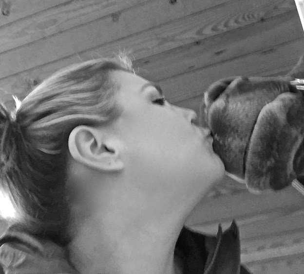
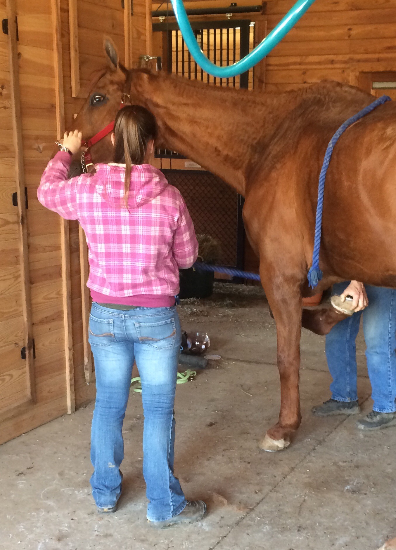
Today Chance got his teeth floated by his very first dentist from 2000!  Due to his cribbing history his front teeth were significantly worn down.  His molars were not in bad shape but were a bit jagged.  The dentist noticed that Chance’s left side was more sensitive to the filing and put a jaw opening device in C’s mouth to keep it open (see below right photo). The molars all looked like they were holding strong and there was no smell that would be indicative of an infection or decay. The dentist indicated that Chance was missing three back molars and that he felt that he was about 24 years old.

The dentist asked me about the nutritional care Chance was receiving due to his age, and I gave him the run down- 2 quarts twice a day of hay stretcher, hay/alfalfa mix throughout the day, 4 quarts of Nutrina Smart Feed Senior twice a day, 2 cups of Rice Bran twice a day in feed, SmartPak Senior Flex and Immune Boost, DuraLactin once a day for arthritic pain and inflammation, Vitamin E once a day, and Transfer Factor for an immune system booster. He continued to explain that when he asks the owners of most of the older horses he goes to sees, they do not have them on the proper diet. I explained that we are still trying to get more weight on Chance but that he has put on a good amount of weight since last summer. He suggested that our next appointment be this December before Chance has the opportunity to go into the winter and lose any weight, which is common in older horses, especially cribbers and thoroughbreds, in the winter months.

Later that day, Chance seemed to have some difficulty eating his hay; wads of hay were scattered around his stall. This is something that I have seen intermittently, maybe once or twice, but not to this extreme.  I decided to give him alfalfa cubes to substitute the hay until the next day when, hopefully, he would be able to eat more easily.  Sure enough the next morning there were no wads of hay!

Therapeutic Shoeing 

By: Christy Corp-Minamiji, DVM

Any discussion of therapeutic shoeing in the horse must begin with a discussion of what therapeutic shoeing is–and what it is not. According to Stephen O’Grady, DVM, MRCVS, professional farrier and owner of Northern Virginia Equine, in Marshall, therapeutic shoeing (or therapeutic farriery) is “the science and art of affecting/influencing the structures of the foot.” It is not a cookie-cutter, “apply shoe A to foot B” magic bullet in the war on equine lameness. To understand how the foot structures can be influenced, we should first have a clear picture of those structures and the forces that act on them.

“In an ideal world, you would begin from a diagnosis,” says Andrew Parks, MA, VetMB, MRCVS, Dipl. ACVS, professor of Large Animal Medicine at the University of Georgia’s College of Veterinary Medicine. “From that diagnosis you would get your treatment goals. By goals I mean the big picture concepts for treatment. You need to have principles (a range of techniques) that allow you to implement your goals.”

O’Grady concurs: “In order to be proficient at therapeutic farriery you must understand the forces that affect the foot. Excess forces or stresses on the hoof capsule lead to deformation, and excess forces or stresses on the internal structures of the foot lead to disease. Everything we are trying to do with therapeutic farriery is to change the forces on the foot.”

For Want of a Foot …

“There is no definition of a perfect foot,” says Parks. What’s considered a “normal” equine foot differs among breeds and disciplines. Thus, when evaluating trimming and shoeing options for a horse, owners and farriers must first consider foot conformation–what you see when you look at a horse standing at rest. Also evaluate limb conformation, which can affect how the horse’s foot bears weight and the way a horse wears a hoof or a shoe. Both limb and hoof conformation might alter the horse’s stride and landing, affecting the forces upon the foot structures. For instance, a club-footed horse’s high-set heels will alter weight distribution, placing more pressure on the toe and heel. While farriery might alter the distribution of the load on the foot, one misconception that O’Grady would like to correct is the idea that you can “change limb conformation by trimming. After 6-8 months (of age), conformation is conformation,” he says. Rather than attempting to make an atypically conformed foot or leg appear “normal,” O’Grady stresses the importance of trimming “for the cards the horse has been dealt.”

Forces affecting the horse hoof

Figure 1: This illustration shows the forces around the center of rotation (the black dot) in a horse at rest. The ground reaction force is transmitted upward through the lamellae to the bony column. The extending force of the coffin joint is balanced by an equal flexing force generated by the deep digital flexor tendon. 

The Foundation: An Appropriate Trim

According to O’Grady, therapeutic shoeing must begin with an appropriately trimmed foot. While much is made of hoof “balance,” O’Grady dislikes the term as he feels it is not clearly defined among practitioners and farriers. He prefers to work from a set of landmarks as clear guidelines to trim each individual foot appropriately:

1) Pick up the foot, and draw a line across the widest part of it. This line will fall just in front of the center of rotation (at the coffin joint). A well-trimmed foot should have approximately even proportions of hoof on either side of that line.
2) Look at the frog. The heels should lie approximately on the same line as the widest part of the frog.
3) Stand the horse on a hard, flat surface so his cannon bone on the leg you’re addressing is perpendicular to the ground. A line drawn down the front of the pastern should be parallel to a line drawn along the dorsal (toe) surface of the hoof wall. This is the hoof-pastern-axis (HPA). A broken-back HPA, in which the front of the foot is angled less steeply than the pastern, shows up in low heels; a broken-forward axis manifests in high heels (club foot).

According to O’Grady, anyone can use these three parameters to assess a horse’s trim. “Trimming is the mainstay of therapeutic shoeing,” he states. Thus, any benefit to be derived from therapeutic shoeing must have a basis in an appropriately trimmed foot. “The first function of a shoe is to protect that which is trimmed and also to complement it,” he says. “Then you can add mechanics … to that trim.”

Physics and the Equine Foot

Newton’s Third Law of Motion states that for every action there is an equal and opposite reaction. Similarly, the support and movement of the equine foot depends upon a system of opposing forces. Understanding these forces is key to understanding the principles of farriery.

In the standing horse:

1) The ground reaction force (GRF, as pictured in Figure 1) is the force transmitted from the ground to the ground surface of the foot, into the hoof wall, and from the wall through the lamellae (interlocking leaflike tissues attaching the hoof to the coffin bone) to the bony column.1 Think of the GRF as the ground pushing up as the foot lands. The GRF extends (i.e., an extensor motion rather than flexor) the coffin and pastern joints and is centered at the center of rotation.2 The GRF is counteracted by two forces.
2) The first of these forces, the weight of the horse, is transmitted down the cannon bone through the fetlock. Since the GRF and the weight of the horse through the cannon bone are both vertical but not aligned, they cause the pastern to rotate so the fetlock becomes closer to the ground.
3) At rest, the horse’s anatomy counters this effect by the upward and backward force of the deep digital flexor tendon (DDFT)–the flexing force illustrated in Figure 1. Picture a string attached to the back of the coffin bone and pulled upward over the navicular bone, over the fetlock, and along the back of the cannon toward the knee.


Transfer Factor: An Answer To Treatment and Prevention of Equine Disease?

by Heather Smith Thomas

For many decades, the typical way veterinarians and horse owners have dealt with disease is by vaccination and by treating sick animals with anti-microbial drugs when signs of illness appear.

By the time the animal shows symptoms, however, damage has already occurred and it can be more difficult to treat the disease. In some instances, irreversible damage has already been done. The use of pathogen-killing drugs is not always as effective as we’d like, and today this use is also being questioned due to the increasing development of drug-resistant pathogens. This microbial resistance diminishes the effectiveness and benefit of some of the drugs we’ve come to rely on.

Horse owners are beginning to look at alternatives to antimicrobial use in dealing with disease. A bright spot in this quest is the use of immune system enhancement and the role of transfer factors. If the immune status of our animals could be enhanced, disease would be less likely to occur, and even if the animals do get sick, the severity and duration of disease could be reduced and they would recover quicker without the need for as much antimicrobial treatment.

Dr. Steve Slagle, a veterinarian in Granite Bay, California, near Sacramento, has been working with a fascinating product that is now available for humans and animals. “The product that I’m using in my practice is a natural immune enhancer and modulator. It derives its efficacy from a protein produced by the immune system’s master immune cells called T lymphocytes. The protein is called transfer factor, and it is also found in cow colostrum. If you buy a bag of dried colostrum (a substitute colostrum product for newborn calves) at the feed store or veterinary supply, about 1% of that product is this protein. We extract that 1% from cow colostrum which enables us to deliver very high levels of transfer factor in our products,” he explains.

“The transfer factors were originally marketed as a human product. I started using them in my veterinary practice in February 1999. So many veterinarians were using the human product that 4Life Research decided to create a veterinary product line for dogs, cats, horses and newborn livestock. Dr. Joe Ramaekers, a colleague of mine, was asked to formulate the product line. Dr. Ramaekers then went on to develop a cancer product for dogs and cats, and a formulation for ruminating livestock,” says Slagle.

“I have been practicing veterinary medicine since 1968 and I have never seen anything that is as exciting as this. I must admit that when I was first informed about transfer factor by a longtime friend, a small animal veterinarian, I was skeptical. He claimed success on so many different types of cases. This didn’t really make sense or seem feasible until I realized some time later that the transfer factors were not treating the particular condition. They were simply enabling the immune system to do its job,” says Slagle

“During the first month I tried it, I used transfer factor on three foals. All three cases were critical and I felt their chances for recovery were slim. The first one was a severe pneumonia. The second was a joint ill infection involving the hock. The third was a terminal septicemia. All three made dramatic recoveries, so I was more than impressed. I was amazed,” he says.

EQUINE PRODUCTS

“The two products from 4Life Research that I use most often in my equine practice are Equine Performance & Show (patented for tumors, EPM, Cushing’s and several other diseases) and Animal Stress Pack (for treating acute conditions). Equine Performance & Show is used primarily as a complete high-end daily supplement with maintenance levels of transfer factor and other immune-enhancing ingredients. I also use it on my chronic cases like tumors, Cushing’s, allergies, and autoimmune diseases like pemphigus (a chronic skin disease). Animal Stress Pack is my emergency treatment, with high levels of transfer factor and other immune enhancers, probiotics, electrolytes and stress vitamins,” says Slagle.

“One of our first studies in horses was done at a major Quarter Horse ranch in Texas that was fighting a losing battle against strangles and a rhodococcus outbreak in which they had already lost several very valuable foals. We put the remaining affected foals on Animal Stress Pack and turned the tide on this very serious situation. All of my infectious disease cases receive the Stress Pack. Even though it is not treating any particular disease, we are dramatically improving the immune response, giving the immune system—which is the ultimate disease fighter—the tools it needs to finish the job,” he explains.

This product is patented for use in horses with EPM. At the latest AAEP Convention in Anaheim, California, Slagle met with Dr. Thomas Bello (a research veterinarian with a private practice, Sandhill Equine Center, in North Carolina). Bello had earlier done the clinical trials for a major drug company on their product for treating EPM.

“The literature on EPM treatments had shown that only between 10% to 20% of horses experience full recovery, returning to their original performance levels. Dr. Bello then became interested in our product and began using it for treating horses with EPM, and getting great results,” says Slagle.

“Dr. Bello then presented a paper at the AVMA convention, which was later published in the Journal of Equine Veterinary Science in 2008—showing that 28 performance horses with EPM were treated with the new EPM product along with the two transfer factor products. At the time of publishing, 82% of those horses were in full recovery. In our recent conversation at the AAEP convention, Dr. Bello told me he now has more than 50 cases in the study, and a recovery rate of over 90%. Apparently the additional immune support was what was needed to bring full recovery. It is also very common to see a relapse in horses that are only being treated with antimicrobials, but Dr. Bello indicated that with his regimen he has not experienced this problem,” says Slagle.

“Since transfer factors are true modulators, my allergic and autoimmune patients go on a daily regimen of transfer factor. Somehow this protein is able to re-educate a confused immune system and bring relief to a large percentage of my equine patients. I generally start them on Performance & Show, along with a week or two of the Stress Pack to front-load the system with high levels of transfer factor. Then when symptoms are under control, we continue with only the Performance & Show,” he explains.

The immune system provides the body with the ability to recognize and remember harmful invaders (pathogenic bacteria, viruses and fungi). Suppressed or damaged immune systems can have disastrous results. One of the most devastating examples is SCID (a genetic defect that occurs in some Arabian foals). They are born without a functioning immune system. After the temporary immunity from the dam’s colostrum is gone, these foals always die of disease.

A healthy immune system has the ability to remember and recognize pathogens, mounting a defense against them. Disease occurs in humans and animals when the immune system is overwhelmed by the pathogen.

HOW IT WORKS

The body’s immune system produces memory molecules whenever it is exposed to disease or receives a vaccination. These memory molecules are bioactive peptides. An example is the “immune” factor passed from a mare to her foal or a cow to her calf via colostrum. This transfer is critical in helping the immune response cells (antibodies) with identification and activation. They are what we might call super boosters in immunity.

Transfer factors were discovered in 1949. Earlier, it had been noticed that immunities could be transferred from one person to another by blood transfusions. In 1949, Dr. H. Sherwood Lawrence, a researcher working on the problem of tuberculosis in humans, found that he could transfer immunity to his patients by using dialyzed leukocytes. When this extract was taken from a blood donor who was resistant to the pathogen and injected into a patient that had no immunity, the immunity of the donor was transferred to the naïve patient. A portion of the lymphocyte (white blood cell) contained what Lawrence dubbed “transfer factor”.

Research was conducted in more than 60 countries (and more than 3500 studies were done) during the 1950s through 1970s and then practically halted. At that point in time, the world’s blood supply was becoming contaminated by HIV and hepatitis C virus and the only known source of transfer factor was blood. Research on this phenomenon was also put on hold because more exciting discoveries revolved around antimicrobial drugs. These were the promising wave of the future that could halt diseases in their tracks. Use of transfer factor was very limited for awhile—especially in veterinary medicine—because it was more expensive to produce than antibiotics. Research did continue, but slowly.

The phenomenon of transfer factor was not actively pursued until the late 1980s when it was discovered that bovine colostrum contains significant amounts of this ingredient that stimulates both aspects of the immune system (humoral and cellular immunity). Veterinary researchers observed a large number of lymphocytic cells in the normal mammary gland secretions of cows, and wondered what role they might play in the health of the newborn calf, realizing that colostrum does more than merely provide passive immune protection. We now know that transfer factor is a lymphokine—one of the protein messengers released by antigen-sensitized lymphocytes (white blood cells).

Chicken eggs also contain transfer factors, and the combination derived from eggs and colostrum increases the effectiveness by 185%. Transfer factors from cow colostrum and eggs are superior to and more functional than transfer factors from humans because animals are exposed to many more species and types of bacteria, viruses and fungi.

As stated by Dr. Richard H. Bennett (Infectious Disease Microbiologist and Immunologist, and former consultant to the National Research Council), transfer factor is one of the most potent messengers in the body and has three effects on the immune system. These are called inducer fractions, antigen specific fractions, and suppressor fractions.

Inducer fractions – One of the functions of transfer factor molecules is to selectively enhance immune surveillance by helping the body recognize various antigens. This selective immune surveillance is made possible by the inducer fractions. One of the veterinarians who consulted with the company that has the patent for extracting transfer factor from colostrum stated that one capsule (200 mg) of transfer factor has the capability of recognizing more than 100,000 different pathogens. Not only can transfer factor be specific for an individual antigen that a lymphocyte might be exposed to, but it can also stimulate a multiple response and provide protection against several strains of that organism.

This enhancement is made possible by the inducer fraction that acts on what are called the Natural Killer (NK) Cells, according to Bennett. The NK cell’s job is to seek out any cells that have been altered by microbes and destroy them. They have a similar protective role in preventing the formation of malignant tumors. The inducer fractions also influence the body’s overall response by increasing the function of the T helper lymphocytes which play a critical role in a balanced immune response to resolve most infections, says Bennett.

The researchers found that they could expose the cow to various bacteria and viruses, and the cow would then produce transfer factor that could stimulate immunity not only to those pathogens but also to other related strains that are much more pathogenic to other species. This is of benefit when using transfer factor to aid disease resistance in horses, for instance. Cows can produce large quantities of colostrum that can then be used for extracting transfer factor that can benefit other species—since transfer factor in horses, cats, dogs, humans and cows has similar structure and identical function.

Another exciting aspect of transfer factor is how quickly the protection is mounted. Immunity from vaccination generally takes 10 to 14 days to develop, whereas transfer factor activates immunity in less than 24 hours.

Antigen specific fractions – Transfer factors act in two ways to “educate” the immune system to respond quickly when confronted by disease threat. One is a response to a specific pathogen such as a cryptosporidium protozoan that might be common to several species, and the other response is to similar pathogens—such as herpes virus infections that differ from one host species to another. Thus transfer factors can “educate” the immune system to recognize and fight a wide array of related, but not identical, infectious agents, according to Bennett.

Suppressor fractions – In every physiological system in the body there are checks and balances, so transfer factor can also act to suppress immune function when necessary. The process of achieving balance is called homeostasis. Once a disease threat has been confronted, and a sufficient response has occurred to thwart it, the body must down-regulate the battle so the immune system can return to a resting state and conserve its resources for the next challenge.

The suppressor fractions signal the T helper lymphocytes and the cytotoxic T cells to slow down their activity and return to a quieter state. This “quieting down” the immune response is important because some pathogenic microbes can hide in certain body tissues and the immune response becomes directed toward those tissues, leading to autoimmune diseases. The suppressor fractions of transfer factor appear to be the way the body limits overzealous immune responses, according to Bennett, and becomes the body’s means to protect itself from an inappropriate immune response.

It seems paradoxical that the transfer factor can both stimulate and suppress immune function, but this is part of its important role. Thus it can prevent autoimmune diseases, and other situations where the body’s own immune response has over-responded to antigens, such as allergic reactions and COPD.

HEALTHIER HORSES

Stressed animals generally become more vulnerable to disease because stress (and the resultant rise in cortisol levels) hinders the immune system. Slagle and Ramaekers tested the transfer factor product on stressed calves to see if it controlled cortisol levels. “We have done two controlled studies on stress and cortisol levels of stressed calves entering the feedlot. One was in Tiffin, Ohio at a private, veterinary-owned and operated feedlot. We repeated that study at Texas A&M. Our results were basically the same. We took blood samples twice daily for 12 days and saw a 46% reduction in cortisol levels in the calves that received transfer factor, with a large decrease in treatments, along with better weight gains,” says Slagle.

“We have not repeated that kind of study in horses, but with the responses (reduced incidence of disease in stress situations) we see in horses, I feel the results would be similar,” he says.

The use of transfer factor to stimulate the body to mount a better immune response to pathogens can reduce the need for antimicrobial drugs. This can help retain their effectiveness longer, since over-use of these drugs has led to increasing numbers of resistant pathogens. We need to find ways to maintain their effectiveness as long as possible.

Transfer factor can boost immunity within a few hours. This makes it very beneficial for use in newborn foals, horses that will be transported, or even as a post-exposure treatment when you know a horse has come into contact with disease agents. Veterinarians have also been using transfer factor to help horses deal with frustrating problems like Cushing’s, laminitis, colitis, cancers, allergies, chronic metritis, EPM, pigeon fever, scours, strangles, and many viral diseases. Helping the immune system help itself is the promising wave of the future.

For more information, Dr. Steve Slagle can be reached in Granite Bay, California at 916-791-2911 or Dr. Joe Ramaekers at 831-476-5050 or check his website: www.ramaekersnutrition.com

“I think I’ll go for a walk outside now- the summer sun is calling my name…”

Sunshine, melting ice and snow, AND 50 degree weather at last! Chance was able to finally enjoy an entire day outside walking around without slipping and sliding ( and without me holding my breath for fear he would fall.)

IMG_2530IMG_2518IMG_2527

Usually, when I arrive at the barn, I will go and see Chance before Lucky to keep him from getting upset.  But today when I got there I didn’t see Chance right away so I decided to bring Lucky in from the paddock.  The next thing I know Chance is standing at the top of the hill staring at me.  I tried to play it cool 🙂 by talking to Chance and continuing to walk Lucky up towards the barn.  That did not work.  Chance freaked out!  He began huffing, Lucky bolted, and Chance refused to walk forward because that would mean Lucky would be out of his range of vision (apparently he thinks this donkey who is maybe up to Chance’s knees in height will attack him).

I finally got Chance to the barn when ice fell off the roof right next to him.  He jolted forward into his stall and all I heard was a bang- Chance hit some part of his body against the stall entrance. I didn’t see any evidence of an injury so I let him calm down for a bit before I began to groom him.  Well, that did not happen….he was pacing and huffing…so I decided to give him some Quietex to help him to calm down and prevent him from hurting himself.  20 minutes later Chance was much calmer.  Although, if you look at the photo, he looks a little drugged.:)

Chance has put on a bit of weight and even though he still needs to gain more, I think he is looking much better.  I began 1 cup of Rice Bran on his food once a day for a week and will up it to 2 cups after.  I am hoping that plus the alfalfa and hay mix, Chance will get some meat on his bones in no time!

The next task, aside from putting on weight and engaging in rehabilitation exercises, is to enhance Chance’s immune system.  Due to his age, medical history, the rehabilitation to help rebuild his neck and hind muscles, and the medications that he has been receiving for EPM, I was advised to add a probiotic to his diet.  The stomach accounts for 60% of a horse’s immune system.  Basically, if the gut is off the chances of a horse contracting a virus, disease, or even injuring himself are much higher.  Not only does the immune system help to fight off illnesses, cancers, etc, it also helps with weight, stress responses, and the effectiveness of treatments.

So, I began to do some research and found some really incredible information which I will talk about in the next post!

Creaking Bones: Information on Equine Arthritis & Prevention

STUdy: Start Measures to Prevent Equine Arthritis Early

“Arthritis is one of the most common reasons we retire horses, and this study shows that prevention of joint damage in early training may be possible through diet,” Coverdale said.

A series of studies by researchers in the department of animal science at Texas A&M University suggest prevention is the best solution to prevent arthritis in young horses.

Josie Coverdale, MS, PhD, associate professor in equine science, and Jessica Lucia, MS, PhD, a former graduate student under Coverdale and now a professor at Sam Houston State University, found that using anti-inflammatory aids mixed with daily feed rations can help decrease joint inflammation in young horses.

“Arthritis is one of the most common reasons we retire horses, and this study shows that prevention of joint damage in early training may be possible through diet,” Coverdale said. “It’s pretty clear the damage comes during early training and that damage often leads to arthritis later in life. A lot of pharmaceuticals are given to treat pain, but few actually help repair the cartilage. We went with the premise that prevention is the best approach rather than trying to treat an existing condition.”

Coverdale said they used the horse production herd at Texas A&M to develop the model and test several diet additives. Lucia read through numerous journals and read a study that used LPS (lipopolysaccharide) injected into the joint for induction of localized inflammation in horses.

“We came across LPS, which has been used in older horses, but not younger horses,” she said. “LPS is the inflammatory part of E. coli, which can be injected using a sterile solution. The beauty of that method is you inject it in the knee and in 24 hours you get pretty quick swelling that is associated with cartilage turnover and related pain.”

This allowed researchers to study the inflammation and breakdown of cartilage over time and mimic the progression of inflammation and cartilage changes associated with intense exercise.

“This initial model study showed us the pattern of inflammation and isolated appropriate markers to measure cartilage breakdown using joint fluid removed from the knee at various time points,” Coverdale said.

Once the LPS model was established to predictably cause joint inflammation, Coverdale said, different dietary strategies were used to try to decrease the amount of inflammation, which included anti-inflammatory dietary supplements such as glucosamine—commonly used by humans to aid in building back damaged cartilage in arthritic joints.

Coverdale said previous data with glucosamine supplementation was “hit or miss with adult horses,” but they wanted to see for themselves and test the theory that prevention in young horses was easier than treating arthritis in the adult.

“We found that it tended to increase new cartilage production and decrease the breakdown of existing cartilage, which was good,” she said.

Study horses received 30 milligrams of glucosamine per kilogram of body weight, Coverdale said.

“We certainly got a positive response, which was what we wanted,” she said.

Another component of Coverdale’s research has been studying conjugated linoleic acid, or CLA, which “is fairly similar to glucosamine in that there are documented anti-inflammatory effects, which may be used to remediate and repair cartilage in joints.”

“Horses receiving supplemental CLA tended to exhibit greater repair of their cartilage when injected with LPS rather than break it down,” she said. “Two percent of the diet was given in the CLA and that can be economically feasible for horse owners.”

The research group has also evaluated horses at varying ages to determine the response to LPS based on age. They concluded that young horses were more likely to synthesize new cartilage in response to inflammation while older horses were more likely to experience cartilage degradation or damage.

“This further illustrates that dietary intervention provided to young horses in training to prevent joint damage may yield the best results,” she said. “With all four of these projects it confirmed that intervening during times of early growth and training with some of these dietary additives is worth it.

“Waiting on down the line as the horse ages is probably too late,” she continued. “Most people are waiting until they see symptoms in these adult horses and by then it is too late. It’s more cost effective and beneficial to do this early. Using it as a prevention method is much better.”

“The initial project to develop the LPS model was funded by the H. Patricia Link Quasi-Endowment funds in the department of animal science,” she said. “This funding was pivotal in furthering the research efforts and helped generate interest from other funding sources such as Cargill Animal Nutrition and the American Quarter Horse Association. The whole premise of the Link funds is to help researchers develop a model or provide preliminary research data to support further funding efforts. This was a perfect example of how this can work.”

16dd9a384ab9509501d38e97d8d8b9bf

For more information select the below link:

Equine Arthritis Information

Vets Urge Horse Owners to Vaccinate Against West Nile Virus

Veterinarians Urge Vaccination against West Nile Virus

By Erica Larson, News Editor

Aug 31, 2012

As of Aug. 21, more than 110 cases of equine West Nile virus (WNV) have been reported this year in the United States, according to the United States Geological Survey. The majority of these affected horses were either not vaccinated against the disease, or not up-to-date on their WNV vaccination.

Since the mosquito season is far from over in some areas of the country, some horse owners might be wondering if a vaccine booster might benefit their animals, or if it’s too late to inoculate a currently unvaccinated horse. To get the answers to these questions, TheHorse.com caught up with Rocky Mason, DVM, an ambulatory equine practitioner from Hagyard Equine Medical Institute in Lexington, Ky.

“Given the recent rise of clinical cases associated with the West Nile virus, I would recommend to owners of unvaccinated horses to consider vaccinating their horses now with a current West Nile vaccine product,” Mason said. “That initial shot needs to be boostered in three to six weeks depending on which commercial product was used. Following that initial two shot series, horses should be vaccinated annually.

“It takes roughly 10 to 14 days for a horse to mount and generate protective antibodies,” he continued. “If the horse is previously unvaccinated it may take two weeks past the second shot to mount protection so the recommendation is to get started now.”

Like with previously unvaccinated horses, Mason recommends starting a new vaccination series in horses with an out-of-date or incomplete vaccination status.

“Horse owners of previously vaccinated horses that have lapsed on their annual vaccine booster will need to go through the initial two-shot series similar to that of unvaccinated horses in order to be in compliance with the commercial product claims,” he explained. “Despite the lapse these horses typically mount an immune response to the first vaccination, but it may not be at a high enough level to prevent viremia, disease and encephalitis. To be safe, the two shot series is recommended in this group as well.”

Mason explained that all commercially available WNV vaccines “have claims for protection of up to a year as determined by challenge models.” That said, even some well vaccinated horses might benefit from a booster this fall, he said.

“More susceptible individuals (young, old, or the immunosuppressed) may require more frequent boostering especially during times of increased exposure like now,” he explained. “It is important to consult with your veterinarian to best determine the needs of your individual horse and its risk assessment. It is very likely given the increased exposure and challenge that the recommendation to booster your horse will be made.”

Finally, Mason stressed that it’s not too late in the year to vaccinate against WNV, even if horses are unvaccinated at present.

“West Nile virus is dependent on mosquitoes for transmission, there are many areas in the country that have insects well into the fall and even winter, necessitating year around immunity. It is important to consult with your veterinarian as only he or she will know when best to time annual boosters in your area.”

Rethinking EPM or Wobbler Diagnoses: Signs of West Nile Virus in Horses

Unfortunately, clinical signs of West Nile virus (WNV) aren’t unique, making it difficult for practitioners to suspect WNV above other neurologic diseases. For this reason, researchers from the University of Florida set out to closely scrutinize the clinical signs of WNV to see if any are particularly unusual. Records of 46 horses admitted to the hospital in 2001 with confirmed cases of WNV encephalitis were reviewed to try to find features that set this disease apart.

The most common clinical signs were weakness and/or ataxia (100% of horses), fever (65%), and muscle fasciculations (twitching; 60%). Fourteen horses (35%) became recumbent from the illness, and 10 of these had to be euthanized. The overall mortality rate was 30%, compared to a rate of 38% across the United States in that year. Importantly, 18 of the 46 horses (39%) had previously received one WNV vaccination, and one horse had received two vaccinations. The most relevant clinical findings were fever, which differentiates WNV from EPM (equine protozoal myeloencephalitis) and cervical myelopathy, and the consistent findings of weakness and/or ataxia, coupled with muscle fasciculations.

Porter, M.B.; Long, M.T.; Getman, L.M.; et al. Journal of the American Veterinary Medical Association, 222 (9), 1241-1247, 2003.

Wobbler Syndrome in Older Horses

Wobbler Syndrome in Older Horses

By Erica Larson, News Editor

Mar 28, 2013

Wobbler Syndrome in Older HorsesThe gold standard for diagnosing CVSM is the meylogram (seen here), a procedure that involves injecting dye into the spinal canal before taking a set of radiographs to evaluate the spinal column’s width and to identify possible sites of compression.

Photo: University of Kentucky Gluck Equine Research Center

The neurologic condition cervical vertebral stenotic myopathy (CVSM, commonly known as wobbler syndrome) is much less common in older horses than it is in young, growing animals. But according to one clinician, this condition should be on all veterinarians’ differential diagnoses list when evaluating an aged horse presenting with neurologic signs and/or neck pain.

At the 2013 Western Veterinary Conference, held Feb. 17-21 in Las Vegas, Nev., Laurie Beard, DVM, MS, Dipl. ACVIM, associate clinical professor at Kansas State University’s College of Veterinary Medicine, presented a review of CVSM in aged horses.

Horses affected by CVSM essentially have a damaged spinal cord. The major causes of spinal cord damage include cervical (neck) vertebrae malformation or trauma.

“The exact pathogenesis of CVSM in older horses is unknown and likely different than younger horses,” Beard said. She said older horses are more likely to have lesions located in the caudal cervical vertebral column (near the base of the neck), between the vertebrae C5 and C6 or C6 and C7. She noted that some studies suggest biomechanical loading related to “wear and tear” could contribute to lesions. Additionally, she said, articular process osteophytosis (bony remodeling) is a common finding in older horses with CVSM.

Beard said common clinical signs associated with CVSM in older horses include:

  • Spinal ataxia (incoordination), generally symmetric with the hind limbs affected more than the front limbs. Beard noted that in some cases, however, mature horses with CVSM might appear more lame than ataxic;
  • Neck pain or stiffness;
  • Neck arthritis; and
  • Decreased appetite.

Beard said veterinarian frequently use standing cervical radiographs to diagnose CVSM in mature horses. However, if cervical radiograph results are inconclusive, a myelogram, considered the gold standard CVSM test, might be required. This procedure involves injecting dye into the spinal canal before taking a set of radiographs to evaluate the spinal column’s width and to identify possible sites of compression.

Treatment options for mature horses diagnosed with CVSM include:

  • Anti-inflammatory drugs (both steroidal and non-steroidal, depending on disease severity);
  • Natural vitamin E supplementation (Beard explained that vitamin E is an antioxidant that could act as an anti-inflammatory in horses with CVSM);
  • Cervical facet steroid injections (this procedure must be repeated every six months or so, but can reduce pain and soft tissue swelling); and
  • Surgical correction (which typically consists of fusing the affected vertebrae–the movement of which causes the spinal cord compression–together using a metal implement called a “basket” in a surgical procedure called cervical stabilization).

The prognosis for for return to work in mature horses with CVSM is variable and depends on the degree of ataxia present, Beard said.

“A horse with mild clinical disease that is a trail riding horse (or low-level performance horse) probably does have a reasonable chance of still performing,” she said. “However, higher level performance horses probably will not be able to perform at the level they were.”

Additionally, horses used for pleasure riding are more likely to have a good prognosis for full return to athletic function than higher performance athletes, she said.

Take-Home Message

Although it’s commonly thought of as a young horse disorder, Beard stressed that veterinarians should consider CVSM as a differential diagnosis in any horse with spinal ataxia. Prognosis for return to work depends on the degree of ataxia present, but many horses show improvement with medical management, she said.

Emerging Neurological Syndromes

Emerging Neurologic Syndromes Discussed at World Equine Vet Meeting

“The more we look for neurologic signs, the more we discover new syndromes,” said I.G. Joe Mayhew, BVSc, FRCVS, PhD, Dipl ACVIM, ECVN, head of Equine Massey and professor of Equine Studies at Massey University in New Zealand, at the 2009 WEVA Congress. Mayhew gave several presentations, including an update on emerging neurologic syndromes.

“Over the last five years or so, we have learned much about existing and new (neurologic) disorders in horses from documentation of careful clinical observations and interventions, and from painstaking pathologic studies with special emphasis on clinicopathologic correlates,” he noted. “This paper will highlight a few of these disorders through which we have added to our understanding of anatomy, physiology, and clinicopathologic correlates–the building blocks for advancing equine neurology.”

Unintentional Parasite

Some nematode parasites that cause neurologic disease in wild and domestic ruminants have now been found to cause problems in horses.

Parelaphostrongylus tenuis is a lungworm that’s life cycle includes cervids (horned animals, such as deer) worldwide, including some in North America. This parasite passes through the host’s central nervous system (CNS) as part of its life cycle. In horses (although not a normal host for the parasite), it has been found to cause acquired cervical torticollis (“wry neck”) due to contraction of the cervical muscles that produce a twisting of the neck and an unnatural posture of the head.

“The scoliosis (curvature of the spine) was clearly argued to be due to loss of afferent cervical proprioceptive inputs because of the dorsal gray column lesions with some white matter involvement accounting for ataxia and weakness,” Mayhew said.

“These nematodes appear to be sensitive to various anthelmintics, such as fenbendazole and ivermectin, and such therapy has been successful when the cases have been treated soon after onset of clinical signs,” he continued.

Cervical Vertebrae Problems

Injury to the cervical vertebrae can affect the horse’s balance. “Special proprioceptive inputs from the cranial cervical vertebral ligaments and muscles pass via at least the C1-3 dorsal spinal nerve roots to ascend the spinal cord via the spinovestibular tract to the caudal vestibular nuclei,” said Mayhew. “These nuclei receive no other afferent inputs. Lesions involving these cranial cervical nerves or the vestibulospinal input to the vestibular apparatus can result in signs of vestibular disease (such as incoordination or loss of balance).”

He said confirmation that apparent neck stiffness and pain, or thoracic limb lameness, is emanating from specific arthritic vertebral articulations “requires radiographic and possibly scintigraphic (on bone scan) evidence of active arthritis and positive relief being achieved from intra- and peri-articular injection of local anesthetic agent.”

Electrodiagnostics

Mayhew reported on a “very sensitive and quite specific electrophysiologic test for disruption of somatic motor pathways in disease states” for horses with neurologic problems such as wobbler syndrome. “When used with the more elaborate, but error-prone, quantitative EMG investigations, this should allow more accurate identification of the presence and location of conduction blocks (electrical impulses to muscles), and, thus, functional lesions, in neurologic disease states such as wobblers and unusual hind limb gait abnormalities,” he explained.

Scandinavian Knuckling Horses

There have been reports of several individual cases and at least five “outbreaks” in groups of horses of a hind-limb knuckling syndrome. In one outbreak 24 cases occurred in a population of 75 animals. Only three of the 24 survived, and one of those three recovered fully.

Veterinarians have described another 75 cases of idiopathic (unknown origin) knuckling in horses in Norway, with no cause determined, but a frequent finding in the cases was poor feed in the form of low-quality baled silage. “Peripheral neurotoxins of plant or nonbiologic origin would be the most likely cause of these crippling syndromes,” said Mayhew.

Equine Motor Neuron Disease

“Acquired equine motor neuron disease (EMND) is a fascinating neuromuscular disorder of horses that does not appear to have existed prior to 1982 and was first described by the late John Cummings (DVM, PhD) and co-workers from Cornell University in 1993,” noted Mayhew. “Hundreds, if not thousands, of horses now have been definitively diagnosed with EMND in North America and from around the world.”

Clinical signs of EMND in horses depend on the stage of the disease, he said. Those signs in early cases often include weight loss in the face of a good to increased appetite, increased recumbency (inability to rise), and slight muscle tremors at rest. “The weight loss often precedes the onset of trembling by several weeks,” he noted. “Many animals display an extended tailhead position that appears to be due to selective involvement of dorsal sacrococcygeal (pertaining to both the sacrum and the coccyx, or the tailbone) muscles that are postural muscles containing a high proportion of Type 1 (slow-contracting muscle) fibers. Atrophy is followed by fibrous contracture leading to an elevated tail position.

“A short-strided gait is commonly seen that can show a rapid placement of the foot at the end of the protraction phase akin to that seen with fibrotic myopathy,” he described. “This also may well be due to fibrous contracture of affected muscles that in this case are caudal thigh muscles involved in stifle flexion and/or hip extension.

“Ophthalmic examination reveals varying degrees of a mosaic pattern with dark brown to yellow brown pigment deposited in the tapetal zone (the tapetum being the iridescent membrane of the choroid of the eye), coupled with a horizontal band of pigment at the junction of the tapetum and nontapetum,” Mayhew said. “A clinical truism for the syndrome is that affected horses move better than they stand.

“Overall study of this disease has given us a better understanding of syndromes of diffuse weakness in horses and particularly weakness involving Type 1 postural, slow-twitch muscles,” he said.

Equine Polysaccharide Storage Myopathy

Equine polysaccharide storage myopathy (EPSM) is an autosomal recessive disorder in Quarter Horse and related breeds and can result in rather exceptional susceptibility to recurrent exertional rhabdomyolysis, reviewed Mayhew.

“The disease EPSM thus refers to the clinical syndrome of muscle disease, particularly rhabdomyolysis, with amylase-resistant, sarcolemmal inclusions of acid mucopolysaccharides evident on muscle biopsy sample,” he said. However, to differentiate EPSM from other diseases of this type, “where there are clinical signs of myopathy (muscle disease or disorder), but histologic evidence of no or mild myopathic changes with excess aggregates or cores of sarcoplasmic (material in which the fibrillae of the muscle fiber are embedded), mostly amylase-sensitive polysaccharide (glycogen), then a distinguishing term such as polysaccharide-associated myopathy should be used.”

EPSM is seen particularly as a likely autosomal recessive trait in Quarter Horses and related breeds and in several other breeds including draft horses.

EPSM is one cause of exertional rhabdomyolysis, and glycogen-associated myopathy probably is also.

“Signs of a hypometric (short-strided) gait, reluctance to move, thoracolumbar lordotic (swayback), and kyphotic (hunchback) postures, and several movement disorders can be seen in association with these disorders,” Mayhew said. He added that “glycogen-associated myopathy is not the cause of most cases of the common postural and movement disorder known as shivers in draft horse and many other breeds.”

Hyperkalemic Periodic Paralysis

Veterinarians have reported the autosomal dominant disease known as hyperkalemic periodic paralysis (HYPP) in Quarter Horse and Quarter Horse-related breeds. Most affected animals are 2 to 3 years old and are male. Homozygous animals (having identical alleles on the paired chromosome) are more severely affected than heterozygotes (those having only one allele).

“The owner notices intermittent episodes of muscle trembling over the body or face, sometimes with intermittent projection of the nictitating membrane (third eyelid), that may lead to involuntary recumbency,” said Mayhew. “Other warning signs include yawning, lowering of the neck, swaying, and disinterest in food and water. During a mild episode the horse is alert, appears distracted and reluctant to move, and may stumble as if weak.” He said that in a full-blown episode, fasciculations (muscle tremors), particularly involving the flank, shoulders, neck, and sometimes the face, progress to staggering, buckling, marked muscle spasms, and paralysis of the limbs might precede involuntary recumbency.

“A severe episode, perhaps following forced exercise, results in severe tremor and tetany (spasming) of many muscles with recumbency and sweating,” he described. “This is followed by a state of flaccidity, possibly with depressed spinal reflexes. Attempts to move the patient result in further tremor and tetany, although the horse remains alert. An episode may last several minutes to hours, typically less than an hour, with full and usually rapid recovery occurring. Between episodes, affected, well-muscled Quarter Horses appear essentially normal.

He said most owners notice stridor (high-pitched respiratory noise) at some time in affected horses. Exercise and rest following exercise might precipitate episodes, which can occur daily or monthly. Stressors such as transportation, weaning, and anesthesia also can trigger episodes.

Stiff Horse Syndrome

Mayhew said a stiff horse syndrome–similar to stiff person syndrome–has been reported. Clinical signs appear to wax and wane and range from mild muscle stiffness to sudden and often violent muscle contractions. Generally, the onset is insidious.

“Between episodes the horse may appear normal, although generalized muscle stiffness may persist,” said Mayhew. “Stiff person syndrome (SPS) has been recognized in humans for some time. It is characterized by muscle rigidity and episodic and often violent muscle cramps.”

In horses, Mayhew described, “Exercise intolerance associated with mild to moderate muscle stiffness may be the only initial clinical sign. This may easily be attributed to a primary myopathy, with pain on muscle palpation, although serum muscle enzyme concentrations remain in the normal range. Components of the syndrome bear resemblance to such disorders as tetanus, equine motor neuron disease, hyperkalemic periodic paralysis, exertional myopathies, and especially the acquired channelopathies associated with the mycotoxicoses, such as perennial ryegrass staggers.

“The most useful diagnostic test is detection of antibodies against the enzyme glutamic acid decarboxylase (GAD) in serum and cerebrospinal fluid, and although some cases have had high anti-GAD titers, several strongly suspected cases have been negative on this test,” Mayhew noted. “It may be necessary to liaise with a human hospital for analyzing for GAD antibodies in the obtained samples. The test relies on cross-reaction with human antigens.

“The overall message really is that with the array of enigmatic movement and postural disorders encountered in equine neurology that appear to be variations on the themes of stringhalt, shivering, and claudication (cramping), a broad approach to delving into possible etiologic mechanisms should be taken that includes the possibility of immune-associated neurotransmitter derangements, such as SPS.”

Grass Sickness

Grass sickness (equine dysautonomia) has been described since the early 20th century, said Mayhew. “Since then it has had quite a devastating effect on equine populations in parts of Western Europe,” he added. “Horses of all breeds, as well as nondomestic equidae and camelids, can be affected, and dogs, cats, rabbits, and hares are affected by similar dysautonomias.”

Mayhew said this disease usually occurs in 3- to 8-year-old horses that are kept outside during late spring and summer, although cases occur year-round. The problem rarely is seen in stalled animals.

“The disease occurs commonly in Northern and Western Europe, particularly in Scotland and England,” he said. “More recently it has been recorded as an epizootic (a disease that appears as new cases in a given animal population, during a given period, at a rate that substantially exceeds what is “expected” based on recent experience) in Hungary, where 15 out of 55 1- to 3-year-old horses in one group succumbed to the disease over one summer, with only three surviving.

“An identical equine dysautonomia known as mal seco occurs in at least Argentina and Chile in South America, and grass sickness appears to now occur in the horse in North America,” stated Mayhew.

Clinical signs can range from acute colic with gastrointestinal stasis (slowing/stopping) and rupture, to anorexia with mild signs of colic and ileus, to chronic intestinal disorder.

“Moderate tachycardia (rapid heart rate), indifference to food, difficulty swallowing, excessive salivation, depressed gastrointestinal sounds, abdominal distension, and usually mild colic are very often present to varying degrees,” noted Mayhew. “Muscular tremor and patchy sweating may be primary signs or may reflect the dehydration, electrolyte imbalances, and colic that occur. Posturing with all feet close together as a weak patient does, ptosis (drooping eyelid), and especially rhinitis sicca (wasting of the mucous membranes and glands with no secretions) are very distinctive signs when present. No definitive clinical diagnostic test exists.”

Atypical Myopathy

Mayhew said several hundred cases of highly fatal, atypical myopathy or myoglobinuria (myoglobin in urine, causing it to appear red-tinged) have been reported in young adult grazing horses. Most of these have been reported in Europe, but they’ve also been detected in North America and Australasia.

“Horses may be found dead or more often showing various signs of reluctance to move, stiff and short strides, apparent sedation, and fine muscle tremors,” he noted. “They quickly become laterally recumbent and urine becomes dark with myoglobin staining, although more subacute cases do occur.”

Symptomatic fluid and analgesic therapy (given as clinical signs dictate) with attentive nursing care for severely ill and often recumbent patients is called for, but the mortality rate of the disease is around 90%.

“Outbreaks do occur, usually in the colder months, and can occur repeatedly on a property,” noted Mayhew. “Access to trees and inclement weather appear to be risk factors for the disease. Plant, bacterial, and fungal toxins have all been considered as possibilities, but the cause or causes remain completely unknown.”

He said preliminary results from one group of investigators suggested thatClostridium sordellii and Clostridium bifermentans toxins might play a role in what they term “pasture myodystrophy.”

Veterinarians with suspected cases are urged to log on to the atypical myopathy alert site (ivis.org) and complete the appropriate forms. This might help in the effort to unravel the epidemiology of this disease.

Lateral Digital Myotenectomy to Treat Stringhalt

Mayhew said stringhalt, also known as springhalt and Hahnentritt (“rooster kick”), is an anciently recorded disease that is characterized by a sudden, apparently involuntary, exaggerated flexion of one or both hind limbs during attempted movement.

“The hind limb motion may be as mild as a slightly excessive flexion to violent movements during which the fetlock or toe will contact the abdomen, thorax, and occasionally the elbow with attempted strides leading to a peculiar bunny hopping and plunging gait,” he described. “The form that usually occurs as outbreaks is seen in Australia, New Zealand, United States, Chile, and Japan, and will be referred to as bilateral, plant-associated stringhalt.”

Usually there is symmetrical or slightly asymmetrical involvement of the pelvic limbs in this syndrome, with prominent distal (farther away from the horse’s core) muscle atrophy in severe cases. The thoracic limbs are also affected in severe cases, with knuckling of the forelimb fetlocks, prominent extension of more proximal joints (those closer to the horse’s body), and atrophy of the distal musculature in association with prominent stringhalt in both hind limbs.

Bilateral stringhalt has been associated with exposure to several plants, notably related species of flat weeds: Hypochoeris radicata, Taraxacum officinale (the common dandelion), and Malva parviflora (mallow weed).

“It is interesting that size and age may be predisposing factors in at least bilateral stringhalt, in so far as older and taller horses tend to become affected in preference to smaller horses, such as ponies and native Chilean breeds,” noted Mayhew. “Although palliative, removing a section of the myotendinous region of the lateral digital extensor muscle relieves the syndrome quite spectacularly in many cases.”

Temporohyoid Osteoarthropathy

Temporohyoid osteoarthropathy (THO) with proliferative osteopathy (bone disease) involving the temporal bone, temporohyoid joint, and hyoid bone in the head, is reported only in adult horses, said Mayhew. It might be subclinical (undetectable) or can result in difficulty chewing or, more often, neurologic syndromes, notably various combinations of facial and vestibulochoclear (ear) nerve dysfunction. (The horse’s tongue lies on the floor of the mouth and is composed of a mass of muscle anchored by the hyoid bone and the bodies of the left and right mandibles–lower jaw.)

“Some of the cases have bilateral disease as determined by endoscopic and radio imaging studies, although the clinical signs are most often unilateral (on one side),” said Mayhew. “The cause of temporohyoid osteoarthropathy is unclear, although to this author a traumatic origin is most plausible in most cases with chronic otitis (ear) media/interna (inflammation of middle/inner ear structures) accounting for a select few cases.

“Regardless of the etiology of the osteoarthritis, clinical signs can occur from either the osteoarthritis itself or from fractures of the adjacent temporal bone and, rarely, basilar bones, due to partial or complete fusion of the joint,” he said. “Physical examination findings may include difficulty chewing, pain on external palpation of the parotid area, headshaking, and behavioral problems–especially when being ridden.

“Once the joint is partly fused, sudden forced head jerking, falling, teeth floating, nasogastric intubation, and sudden prolonged vocalization can cause periarticular fractures of the petrous temporal bone, resulting in combinations of an abrupt onset of facial and vestibular nerve dysfunction,” he noted. “Endoscopic examination of the guttural pouch is probably superior to plain radiographic imaging in confirming the presence of the disease by revealing enlargement of the proximal stylohyoid bone due to osteoarthritis when compared to the opposite side. “In acute or progressive cases having ill-defined endoscopic and plain radiographic imaging findings, gamma scintigraphy should be considered as a diagnostic aid.”

Mayhew said he was aware of several cases that improved over time, only to show further signs relative to facial and vestibular nerve dysfunction in weeks to months time. “These would seem to be ideal candidates for unilateral surgical disunion of the hyoid apparatus,” he noted. “Initial surgical disunion of the hyoid apparatus was performed by removal of a midshaft portion of the stylohyoid bone. To reduce the temporary difficulties in swallowing encountered and to reduce the possibility of other real and potential complications of this surgery, the technique of ceratohyoidectomy was proposed and used with success.”

He said that except for major cranial fractures and residual eye problems, the outlook for survival with residual neurologic deficits is quite good. “Of 33 cases of temporohyoid osteoarthropathy, 20 cases survived for which there were longer term follow-up details,” he reported. “Of these, 70% returned to previous level of use, although more than 50% of the 20 horses still had evidence of facial nerve deficits and/or vestibular dysfunction.

“Thus, in spite of some optimistic suggestions, if full athletic performance without neurologic dysfunction is required, then the prognosis with or without surgical intervention has to be fair to guarded for these cases,” he said. “Cases of THO have given us a better insight into the ability of horses to accommodate to vestibular dysfunction and to survive with degrees of facial paralysis.”

Post-Anesthetic Cerebral Necrosis

A newly defined, unexpected complication of apparently routine general anesthesia in some mature horses is diffuse and severe cerebral necrosis, resulting in signs of diffuse (not concentrated or localized) encephalopathy immediately or some hours to days after recovery from anesthesia, reported Mayhew.

There is cerebral edema (fluid swelling) and laminar neuronal cortical necrosis associated with generalized signs that predominantly consist of somnolence (drowsiness) to dementia, central blindness, wandering compulsively, pushing against objects, and ataxia.

“One patient with this tentative diagnosis that recovered showed prominent muzzle and ear twitching, very reminiscent of patients suffering from bacterial meningitis and from West Nile viral meningoencephalitis (inflammation of the brain and the meninges–the membranes that cover the brain),” he said.

Early Detection of Equine Arthritis?

Is Early Detection of Arthritis in Horses Finally a Reality?

Is Early Detection of Arthritis in Horses Finally a Reality?Radiography’s ability to correctly identify joints without OA was 97%, meaning it had few false-positives, and that radiography was equal to or better than MRI for detecting early joint changes consistent with OA.

Photo: Kevin Thompson/The Horse

Osteoarthritis (OA) is a progressive deterioration of joint health with no known cure. Not only does OA negatively affect athleticism and quality of life but it is also a major cause of economic loss throughout the equine industry.

For years researchers have been trying to find ways to diagnose OA early in the course of disease to either slow or, better yet, arrest its progression. And although OA has proven a stubborn opponent, an international group of researchers recently found that radiographs (X rays) and low-field MRI appear to be useful tools for diagnosing OA.

“For our study we chose to use Icelandic horses, a breed that is known to have a high prevalence of OA and one in which a large number of older riding horses are culled due to the pain and lameness that result from the disease,” explained Charles Ley, BVSc, Dipl. ECVDI, PhD, from the Swedish University of Agricultural Sciences, in Uppsala. “Young horses without obvious lameness were used in the study in order to include horses likely to have a very early stage of the disease and normal horses. We chose to use two noninvasive and clinically available imaging techniques—radiography and MRI—to see if it was possible to detect early OA changes in the joints.”

Ley and colleagues collected 75 hock joint radiographs and MRIs from 38 Icelandic horses between the ages of 27 and 31 months. The team then used microscopy to classify joints as positive or negative for OA.

The team classified 42 of the 75 joints as OA-positive after they detected lesions on both radiography and MRI that corresponded with OA, including mineralization front defects and joint margin lesions. The team determined that radiography’s ability to correctly identify joints without OA was 97%, meaning it had few false-positives, and that radiography was equal to or better than MRI for detecting early joint changes consistent with OA.

“Radiography is a widely available, cost-effective, and repeatable method, and the high specificity and high frequency of the detection of mineralization front defects in radiographs suggests that this is a promising marker of early OA in the distal intertarsal joint (one of the middle hock joints),” Ley concluded. “Such a tool has a vital role in selecting horses for inclusion in long-term studies of how and why OA develops and evaluating early intervention and prevention methods for OA.”

The study, “Detection of early osteoarthritis in the centrodistal joints of Icelandic horses: evaluation of radiography and low-field magnetic resonance imaging,” will appear in an upcoming issue of the Equine Veterinary Journal.