Category Archives: Prevention

Trust Your Instincts

Chance being lunged after many years of not being lunged due to a DDFT injury and possible EPM treatments.  He is still stiff and needs to build up the muscle in his hind end thus why he is being lunged.  In addition to his chiropractic adjustments, acupuncture, stretches and massages, and all natural pain medication (MicroLactin), he is also doing physical therapy- walking over poles, walking serpentines, and walking up and down hills.

It Just Keeps Getting Better & Better

Two days ago Chance’s vet came out to do a follow up and to give him and Luck their Spring shots.

Chance got some chiropractic adjustments and acupuncture as well. The vet stated that Chance was showing improvements in his Cervical (neck) flexibility and still had some tightness on his hind-end. She did one new stretch with him which entailed her lifting his front leg while her assistant had him bend his neck to the opposite side. He was able to do it on both sides while remaining balanced!!!! Where as before he could barely do cervical stretch with all four legs on the ground!

IMG_3031

IMG_3030  

The vet said that Chance has gained weight and could use another 50-75lbs. She asked if he is finishing his dinner (4q twice a day) to which I answered yes. She suggested upping his feed to another quart twice a day totaling 5 quarts twice a day.

Chance’s feed schedule now includes the following:

AM: 5 q of Safe Choice Senior Feed by Nutrina, Transfer Factor (immune system booster), MicroLactin (for pain and inflammation). Hay Alfalfa mix, and 2q of Hay Stretcher

PM: 5 q of SCSF, SmartPak (Senior Flex supplement, immune system enhancer),  Hay/alfalfa mix, and 2 q of Hay Stretcher.

Lucky got his first round of shots today and he was so well behaved! He stood there calmly and put his head under my arm while he got his shots.

The vet also took a look at Luck’s teeth.

There are four ways to age an equine by his teeth:

  • Occurrence of permanent teeth
  • Disappearance of cups
  • Angle of incidence
  • Shape of the surface of the teeth

Well, Luck still has two baby teeth which do not seem to have adult teeth behind them that would ordinarily push out the baby teeth. So there goes option 1. 

His teeth no longer have cups on them and are completely smooth which indicates he is around age 10/11. 

I, along with Luck’s most recent owner, thought he was about 5 years old. However, his teeth seem to tell a different story. I’m wondering if the fact that Luck still has two of his baby teeth could be the reason for the cups prematurely disappearing? Or if he really is 10/11 years old….guess it’s time for a dental appointment. 

Hannibal. 

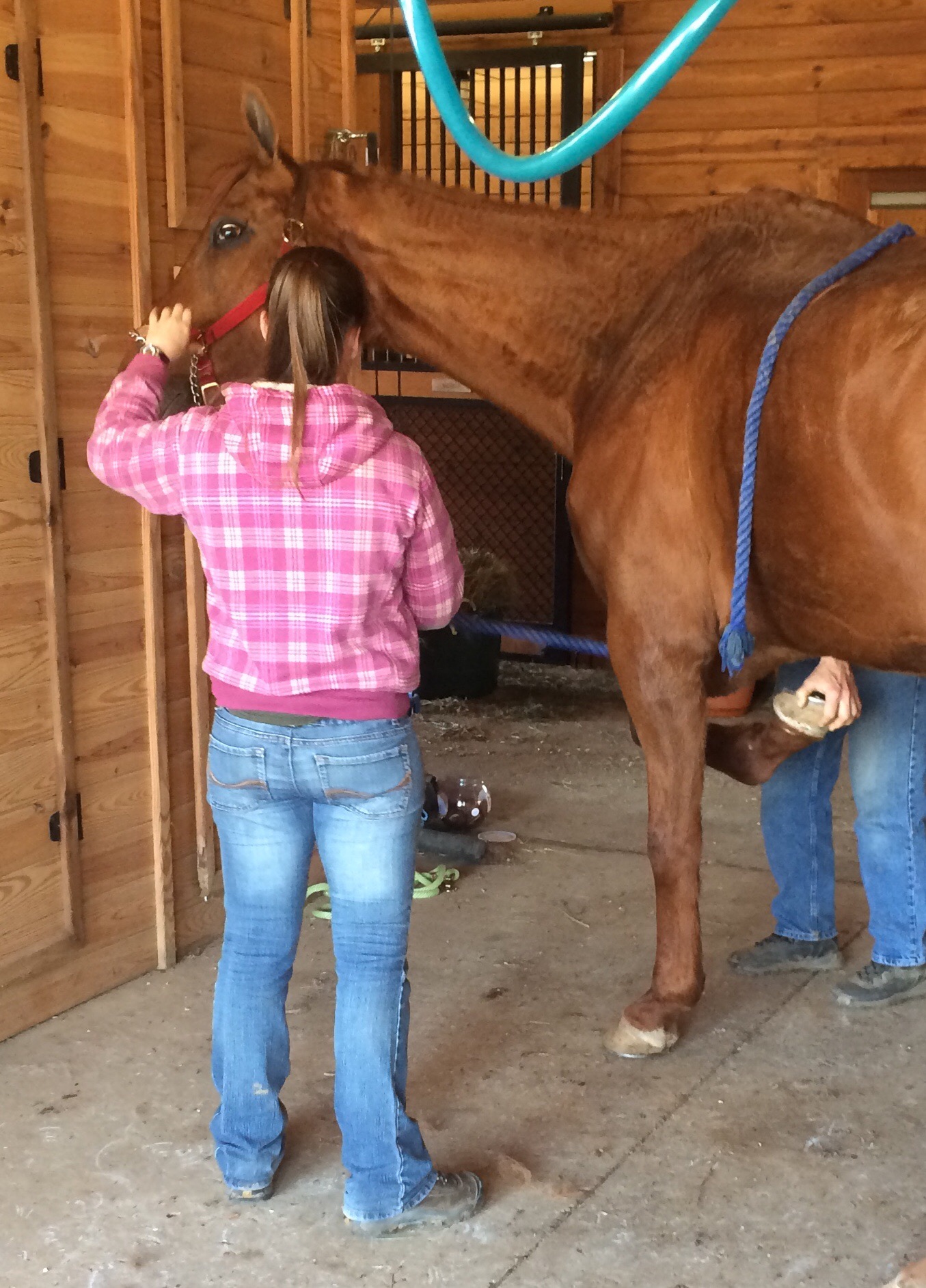
Today Chance got his teeth floated by his very first dentist from 2000!  Due to his cribbing history his front teeth were significantly worn down.  His molars were not in bad shape but were a bit jagged.  The dentist noticed that Chance’s left side was more sensitive to the filing and put a jaw opening device in C’s mouth to keep it open (see below right photo). The molars all looked like they were holding strong and there was no smell that would be indicative of an infection or decay. The dentist indicated that Chance was missing three back molars and that he felt that he was about 24 years old.

The dentist asked me about the nutritional care Chance was receiving due to his age, and I gave him the run down- 2 quarts twice a day of hay stretcher, hay/alfalfa mix throughout the day, 4 quarts of Nutrina Smart Feed Senior twice a day, 2 cups of Rice Bran twice a day in feed, SmartPak Senior Flex and Immune Boost, DuraLactin once a day for arthritic pain and inflammation, Vitamin E once a day, and Transfer Factor for an immune system booster. He continued to explain that when he asks the owners of most of the older horses he goes to sees, they do not have them on the proper diet. I explained that we are still trying to get more weight on Chance but that he has put on a good amount of weight since last summer. He suggested that our next appointment be this December before Chance has the opportunity to go into the winter and lose any weight, which is common in older horses, especially cribbers and thoroughbreds, in the winter months.

Later that day, Chance seemed to have some difficulty eating his hay; wads of hay were scattered around his stall. This is something that I have seen intermittently, maybe once or twice, but not to this extreme.  I decided to give him alfalfa cubes to substitute the hay until the next day when, hopefully, he would be able to eat more easily.  Sure enough the next morning there were no wads of hay!

Transfer Factor: An Answer To Treatment and Prevention of Equine Disease?

by Heather Smith Thomas

For many decades, the typical way veterinarians and horse owners have dealt with disease is by vaccination and by treating sick animals with anti-microbial drugs when signs of illness appear.

By the time the animal shows symptoms, however, damage has already occurred and it can be more difficult to treat the disease. In some instances, irreversible damage has already been done. The use of pathogen-killing drugs is not always as effective as we’d like, and today this use is also being questioned due to the increasing development of drug-resistant pathogens. This microbial resistance diminishes the effectiveness and benefit of some of the drugs we’ve come to rely on.

Horse owners are beginning to look at alternatives to antimicrobial use in dealing with disease. A bright spot in this quest is the use of immune system enhancement and the role of transfer factors. If the immune status of our animals could be enhanced, disease would be less likely to occur, and even if the animals do get sick, the severity and duration of disease could be reduced and they would recover quicker without the need for as much antimicrobial treatment.

Dr. Steve Slagle, a veterinarian in Granite Bay, California, near Sacramento, has been working with a fascinating product that is now available for humans and animals. “The product that I’m using in my practice is a natural immune enhancer and modulator. It derives its efficacy from a protein produced by the immune system’s master immune cells called T lymphocytes. The protein is called transfer factor, and it is also found in cow colostrum. If you buy a bag of dried colostrum (a substitute colostrum product for newborn calves) at the feed store or veterinary supply, about 1% of that product is this protein. We extract that 1% from cow colostrum which enables us to deliver very high levels of transfer factor in our products,” he explains.

“The transfer factors were originally marketed as a human product. I started using them in my veterinary practice in February 1999. So many veterinarians were using the human product that 4Life Research decided to create a veterinary product line for dogs, cats, horses and newborn livestock. Dr. Joe Ramaekers, a colleague of mine, was asked to formulate the product line. Dr. Ramaekers then went on to develop a cancer product for dogs and cats, and a formulation for ruminating livestock,” says Slagle.

“I have been practicing veterinary medicine since 1968 and I have never seen anything that is as exciting as this. I must admit that when I was first informed about transfer factor by a longtime friend, a small animal veterinarian, I was skeptical. He claimed success on so many different types of cases. This didn’t really make sense or seem feasible until I realized some time later that the transfer factors were not treating the particular condition. They were simply enabling the immune system to do its job,” says Slagle

“During the first month I tried it, I used transfer factor on three foals. All three cases were critical and I felt their chances for recovery were slim. The first one was a severe pneumonia. The second was a joint ill infection involving the hock. The third was a terminal septicemia. All three made dramatic recoveries, so I was more than impressed. I was amazed,” he says.

EQUINE PRODUCTS

“The two products from 4Life Research that I use most often in my equine practice are Equine Performance & Show (patented for tumors, EPM, Cushing’s and several other diseases) and Animal Stress Pack (for treating acute conditions). Equine Performance & Show is used primarily as a complete high-end daily supplement with maintenance levels of transfer factor and other immune-enhancing ingredients. I also use it on my chronic cases like tumors, Cushing’s, allergies, and autoimmune diseases like pemphigus (a chronic skin disease). Animal Stress Pack is my emergency treatment, with high levels of transfer factor and other immune enhancers, probiotics, electrolytes and stress vitamins,” says Slagle.

“One of our first studies in horses was done at a major Quarter Horse ranch in Texas that was fighting a losing battle against strangles and a rhodococcus outbreak in which they had already lost several very valuable foals. We put the remaining affected foals on Animal Stress Pack and turned the tide on this very serious situation. All of my infectious disease cases receive the Stress Pack. Even though it is not treating any particular disease, we are dramatically improving the immune response, giving the immune system—which is the ultimate disease fighter—the tools it needs to finish the job,” he explains.

This product is patented for use in horses with EPM. At the latest AAEP Convention in Anaheim, California, Slagle met with Dr. Thomas Bello (a research veterinarian with a private practice, Sandhill Equine Center, in North Carolina). Bello had earlier done the clinical trials for a major drug company on their product for treating EPM.

“The literature on EPM treatments had shown that only between 10% to 20% of horses experience full recovery, returning to their original performance levels. Dr. Bello then became interested in our product and began using it for treating horses with EPM, and getting great results,” says Slagle.

“Dr. Bello then presented a paper at the AVMA convention, which was later published in the Journal of Equine Veterinary Science in 2008—showing that 28 performance horses with EPM were treated with the new EPM product along with the two transfer factor products. At the time of publishing, 82% of those horses were in full recovery. In our recent conversation at the AAEP convention, Dr. Bello told me he now has more than 50 cases in the study, and a recovery rate of over 90%. Apparently the additional immune support was what was needed to bring full recovery. It is also very common to see a relapse in horses that are only being treated with antimicrobials, but Dr. Bello indicated that with his regimen he has not experienced this problem,” says Slagle.

“Since transfer factors are true modulators, my allergic and autoimmune patients go on a daily regimen of transfer factor. Somehow this protein is able to re-educate a confused immune system and bring relief to a large percentage of my equine patients. I generally start them on Performance & Show, along with a week or two of the Stress Pack to front-load the system with high levels of transfer factor. Then when symptoms are under control, we continue with only the Performance & Show,” he explains.

The immune system provides the body with the ability to recognize and remember harmful invaders (pathogenic bacteria, viruses and fungi). Suppressed or damaged immune systems can have disastrous results. One of the most devastating examples is SCID (a genetic defect that occurs in some Arabian foals). They are born without a functioning immune system. After the temporary immunity from the dam’s colostrum is gone, these foals always die of disease.

A healthy immune system has the ability to remember and recognize pathogens, mounting a defense against them. Disease occurs in humans and animals when the immune system is overwhelmed by the pathogen.

HOW IT WORKS

The body’s immune system produces memory molecules whenever it is exposed to disease or receives a vaccination. These memory molecules are bioactive peptides. An example is the “immune” factor passed from a mare to her foal or a cow to her calf via colostrum. This transfer is critical in helping the immune response cells (antibodies) with identification and activation. They are what we might call super boosters in immunity.

Transfer factors were discovered in 1949. Earlier, it had been noticed that immunities could be transferred from one person to another by blood transfusions. In 1949, Dr. H. Sherwood Lawrence, a researcher working on the problem of tuberculosis in humans, found that he could transfer immunity to his patients by using dialyzed leukocytes. When this extract was taken from a blood donor who was resistant to the pathogen and injected into a patient that had no immunity, the immunity of the donor was transferred to the naïve patient. A portion of the lymphocyte (white blood cell) contained what Lawrence dubbed “transfer factor”.

Research was conducted in more than 60 countries (and more than 3500 studies were done) during the 1950s through 1970s and then practically halted. At that point in time, the world’s blood supply was becoming contaminated by HIV and hepatitis C virus and the only known source of transfer factor was blood. Research on this phenomenon was also put on hold because more exciting discoveries revolved around antimicrobial drugs. These were the promising wave of the future that could halt diseases in their tracks. Use of transfer factor was very limited for awhile—especially in veterinary medicine—because it was more expensive to produce than antibiotics. Research did continue, but slowly.

The phenomenon of transfer factor was not actively pursued until the late 1980s when it was discovered that bovine colostrum contains significant amounts of this ingredient that stimulates both aspects of the immune system (humoral and cellular immunity). Veterinary researchers observed a large number of lymphocytic cells in the normal mammary gland secretions of cows, and wondered what role they might play in the health of the newborn calf, realizing that colostrum does more than merely provide passive immune protection. We now know that transfer factor is a lymphokine—one of the protein messengers released by antigen-sensitized lymphocytes (white blood cells).

Chicken eggs also contain transfer factors, and the combination derived from eggs and colostrum increases the effectiveness by 185%. Transfer factors from cow colostrum and eggs are superior to and more functional than transfer factors from humans because animals are exposed to many more species and types of bacteria, viruses and fungi.

As stated by Dr. Richard H. Bennett (Infectious Disease Microbiologist and Immunologist, and former consultant to the National Research Council), transfer factor is one of the most potent messengers in the body and has three effects on the immune system. These are called inducer fractions, antigen specific fractions, and suppressor fractions.

Inducer fractions – One of the functions of transfer factor molecules is to selectively enhance immune surveillance by helping the body recognize various antigens. This selective immune surveillance is made possible by the inducer fractions. One of the veterinarians who consulted with the company that has the patent for extracting transfer factor from colostrum stated that one capsule (200 mg) of transfer factor has the capability of recognizing more than 100,000 different pathogens. Not only can transfer factor be specific for an individual antigen that a lymphocyte might be exposed to, but it can also stimulate a multiple response and provide protection against several strains of that organism.

This enhancement is made possible by the inducer fraction that acts on what are called the Natural Killer (NK) Cells, according to Bennett. The NK cell’s job is to seek out any cells that have been altered by microbes and destroy them. They have a similar protective role in preventing the formation of malignant tumors. The inducer fractions also influence the body’s overall response by increasing the function of the T helper lymphocytes which play a critical role in a balanced immune response to resolve most infections, says Bennett.

The researchers found that they could expose the cow to various bacteria and viruses, and the cow would then produce transfer factor that could stimulate immunity not only to those pathogens but also to other related strains that are much more pathogenic to other species. This is of benefit when using transfer factor to aid disease resistance in horses, for instance. Cows can produce large quantities of colostrum that can then be used for extracting transfer factor that can benefit other species—since transfer factor in horses, cats, dogs, humans and cows has similar structure and identical function.

Another exciting aspect of transfer factor is how quickly the protection is mounted. Immunity from vaccination generally takes 10 to 14 days to develop, whereas transfer factor activates immunity in less than 24 hours.

Antigen specific fractions – Transfer factors act in two ways to “educate” the immune system to respond quickly when confronted by disease threat. One is a response to a specific pathogen such as a cryptosporidium protozoan that might be common to several species, and the other response is to similar pathogens—such as herpes virus infections that differ from one host species to another. Thus transfer factors can “educate” the immune system to recognize and fight a wide array of related, but not identical, infectious agents, according to Bennett.

Suppressor fractions – In every physiological system in the body there are checks and balances, so transfer factor can also act to suppress immune function when necessary. The process of achieving balance is called homeostasis. Once a disease threat has been confronted, and a sufficient response has occurred to thwart it, the body must down-regulate the battle so the immune system can return to a resting state and conserve its resources for the next challenge.

The suppressor fractions signal the T helper lymphocytes and the cytotoxic T cells to slow down their activity and return to a quieter state. This “quieting down” the immune response is important because some pathogenic microbes can hide in certain body tissues and the immune response becomes directed toward those tissues, leading to autoimmune diseases. The suppressor fractions of transfer factor appear to be the way the body limits overzealous immune responses, according to Bennett, and becomes the body’s means to protect itself from an inappropriate immune response.

It seems paradoxical that the transfer factor can both stimulate and suppress immune function, but this is part of its important role. Thus it can prevent autoimmune diseases, and other situations where the body’s own immune response has over-responded to antigens, such as allergic reactions and COPD.

HEALTHIER HORSES

Stressed animals generally become more vulnerable to disease because stress (and the resultant rise in cortisol levels) hinders the immune system. Slagle and Ramaekers tested the transfer factor product on stressed calves to see if it controlled cortisol levels. “We have done two controlled studies on stress and cortisol levels of stressed calves entering the feedlot. One was in Tiffin, Ohio at a private, veterinary-owned and operated feedlot. We repeated that study at Texas A&M. Our results were basically the same. We took blood samples twice daily for 12 days and saw a 46% reduction in cortisol levels in the calves that received transfer factor, with a large decrease in treatments, along with better weight gains,” says Slagle.

“We have not repeated that kind of study in horses, but with the responses (reduced incidence of disease in stress situations) we see in horses, I feel the results would be similar,” he says.

The use of transfer factor to stimulate the body to mount a better immune response to pathogens can reduce the need for antimicrobial drugs. This can help retain their effectiveness longer, since over-use of these drugs has led to increasing numbers of resistant pathogens. We need to find ways to maintain their effectiveness as long as possible.

Transfer factor can boost immunity within a few hours. This makes it very beneficial for use in newborn foals, horses that will be transported, or even as a post-exposure treatment when you know a horse has come into contact with disease agents. Veterinarians have also been using transfer factor to help horses deal with frustrating problems like Cushing’s, laminitis, colitis, cancers, allergies, chronic metritis, EPM, pigeon fever, scours, strangles, and many viral diseases. Helping the immune system help itself is the promising wave of the future.

For more information, Dr. Steve Slagle can be reached in Granite Bay, California at 916-791-2911 or Dr. Joe Ramaekers at 831-476-5050 or check his website: www.ramaekersnutrition.com

How To Prepare For An Equine Emergency

Be Prepared for an Equine Emergency

You don’t want to waste time in an equine emergency! The American Association of Equine Practitioners (AAEP) offers these tips to keep you organized and calm in your horse’s time of need.


If you own horses long enough, sooner or later you are likely to confront a medical emergency. From lacerations to colic to foaling difficulties, there are many emergencies that a horse owner may encounter. You must know how to recognize serious problems and respond promptly, taking appropriate action while awaiting the arrival of your veterinarian.

Preparation is vital when confronted with a medical emergency. No matter the situation you may face, mentally rehearse the steps you will take to avoid letting panic take control. Follow these guidelines from the American Association of Equine Practitioners (AAEP) to help you prepare for an equine emergency:

    1. Keep your veterinarian’s number by each phone, including how the practitioner can be reached after hours.
    1. Consult with your regular veterinarian regarding a back-up or referring veterinarian’s number in case you cannot reach your regular veterinarian quickly enough.
    1. Know in advance the most direct route to an equine surgery center in case you need to transport the horse.
    1. Post the names and phone numbers of nearby friends and neighbors who can assist you in an emergency while you wait for the veterinarian.
    1. Prepare a first aid kit and store it in a clean, dry, readily accessible place. Make sure that family members and other barn users know where the kit is. Also keep a first aid kit in your horse trailer or towing vehicle, and a pared-down version to carry on the trail.First aid kits can be simple or elaborate. Here is a short list of essential items:
        • Cotton roll
        • Cling wrap
        • Gauze pads, in assorted sizes
        • Sharp scissors
        • Cup or container
        • Rectal thermometer with string and clip attached
        • Surgical scrub and antiseptic solution
        • Latex gloves
        • Saline solution
        • Stethoscope
        • Clippers

Many accidents can be prevented by taking the time to evaluate your horse’s environment and removing potential hazards. Mentally rehearse your emergency action plan. In an emergency, time is critical. Don’t be concerned with overreacting or annoying your veterinarian. By acting quickly and promptly, you can minimize the consequences of an injury or illness.

For more information about emergency care, ask your equine veterinarian for the “Emergency Care” brochure, provided by the AAEP in partnership with Bayer Corporation, Animal Health. More information can also be obtained by visiting the AAEP’s horse health web site, www.myHorseMatters.com.

The American Association of Equine Practitioners, headquartered in Lexington, Kentucky, was founded in 1954 as a non-profit organization dedicated to the health and welfare of the horse.

– See more at: http://practicalhorsemanmag.com/article/eqemergenc2576#sthash.YFzhhSOX.dpuf

A Month Later

Luckily, after about one week of stall rest and hand walking, along with a stronger anti-inflammatory, Chance has begun to show improvements over the last couple weeks of treatment.

Chance has almost completed his first 30 days of EPM treatment and has about a day or so left of the Protazil. He is going to continue his other medications and supplements:

1. SmartPak: senior flex and immune boost
2. Vitamin E
3. MicroLactin (amazing) to help with regrowth of his cells, inflammation, and pain.

As I’m doing research, and trying to come up with a plan of action, once again I am inundated with opinions…medication, exercise, holistic, massage, acupuncture, chiropractic, organic, shoeing, etc.

I know I need to continue therapy, or start a new therapy. But which one do I choose?

Do I go the holistic approach and work with an acupuncturist, chiropractor, massage therapist, and vitamins such as Vita Royals? Do I go organic? Or do I try Marquis? Another month of Protazil? Oraquin-10? Rebalance has been linked to a number of recent deaths in the past handful of months. If I go the organic or holistic approach do I run the risk of the disease progressing? If I go with the medication do I run the risk of yet another “treatment crisis”?

I contacted Vet4 and he suggested doing another round of the Protazil or Marquis.  I decided on trying Marquis and waited for it to arrive.

Chance after a month on Protazil 

CHRONIC PROGRESSIVE LYMPHEDEMA (CPL) due to Scratches

Chance has always been fighting “scratches” on his back legs.  Frustrating, painful, and never seem to completely go away.  Could scratches have caused this?  My thoughts- scratches allowed bacteria to enter the leg, the infection settled on the DDFT sheath and caused the current flare up.  Below is some research I found on possible conditions due to scratches that caused similar symptoms Chance had been experiencing.

CHRONIC PROGRESSIVE LYMPHEDEMA (CPL) due to Scratches

A condition characterized by progressive swelling, hyperkeratosis and fibrosis of distal limbs has been characterized in Shires, Clydesdales and Belgian Draft horses and unfortunately affects numerous horses within these breeds. The disease has also been recognized in Gipsy Vanners; however, only a few horses have been evaluated at this point of time. This chronic progressive disease starts at an early age, progresses throughout the life of the horse and often ends in disfigurement and disability of the legs, which inevitably leads to the horse’s premature death. The pathologic changes and clinical signs closely resemble a condition known in humans as chronic lymphedema or elephantiasis nostras verrucosa. The condition has therefore been referred to as chronic progressive lymphedema (CPL). The lower leg swelling is caused by abnormal functioning of the lymphatic system in the skin, which results in chronic lymphedema (swelling), fibrosis, decreased perfusion, a compromised immune system and subsequent secondary infections of the skin.

The clinical signs of this disease are highly variable. It is often first addressed as a marked and “therapy-resistant” pastern dermatitis (scratches). The earliest lesions, however, are characterized by skin thickening, slight crusting and possible skin folds in the pastern area. While readily palpable, these early lesions are often not appreciated visually as the heavy feathering in these breeds covers these areas. Upon clipping of the lower legs, it becomes obvious that the lesions are far more extensive than expected. Secondary infections develop very easily in these horse’s legs and usually consist of chorioptic mange and/or bacterial infections. Pigmented and non-pigmented skin of the lower legs are affected. Appropriate treatment of the infections (pastern dermatitis) is not successful as underlying poor perfusion, lymphedema and hyperkeratosis in association with the heavy feathering present perfect conditions for repetitive infections with both chorioptic mange as well as bacterial infections. Recurrent infections and inflammation will enhance the lymphedema and hence, the condition becomes more chronic. As a result, the lower leg enlargement becomes permanent and the swelling firm on palpation. More thick skin folds and large, poorly defined, firm nodules develop. The nodules may become quite large and often are described as “golf ball” or even “baseball” in size. Both skin folds and nodules first develop in the back of the pastern area. With progression, they may extend and encircle the entire lower leg. The nodules become a mechanical problem because they interfere with free movement and frequently are injured during exercise. This disease often progresses to include massive secondary infections that produce copious amounts of foul-smelling exudates, generalized illness, debilitation and even death.

TREATMENT/MANAGEMENT

Please keep in mind that none of these treatments listed below will “heal” chronic progressive lymphedema (CPL). However, a rigorous management following our suggestions below will assist you to slow down the process and even make some of the nodular lesions disappear. Your horse will need this management the rest of its life.

• Clipping of the feathers
Long and dense feathering makes management of lymphedema more difficult. We highly recommend clipping the feathers and keep them short, if horses are not presented at shows. If you have a show horse, we still recommend to clip the feathers to initiate a rigorous treatment. As the skin condition improves and the edema is reducing – you may have a better chance to keep the horse’s legs in better condition by. careful repetitive treatment, while the feathering is growing back. The feathers are usually back to their original length in about 10-12 months.

• Treatment of skin infections
Progression of lymphedema is also associated with deposition of fibrous tissue and formation of fibrotic nodules.. As a result, these horses have a poor blood circulation and immune response in the skin of their legs. They tend to built up a thick keratin layer. The long feathering further occludes the skin surface, which then remains humid. These factors provide the perfect culture environment for infectious pathogens. This explains why horses with CPL constantly battle recurrent infections with mites (Chorioptic mange) and bacterial infections (Staphylococcus, Dermatophilus).

Horses with CPL should consistently be treated against reinfestation of mites and bacteria:

Topical treatments:

• Careful washing, cleaning and drying of the legs on a routine basis is essential. Horses with long feathering may require blow-drying of their legs. We recommend using a product manufactured by HydroSurge Inc. ( http://www.hydrosurge.com ) called Apricot Sulfur Skin Treatment Shampoo.

• Frontline spray to treat chorioptic mange (do not use Frontline on pregnant and nursing mares)

• The best and most economical topical treatment is to find a source of wettable sulfur powder (“flowers of sulfur”). This can usually be found through a vineyard supply or at your local nursery (certain “rose dust” preparations). Mix this powder with mineral oil in to form a creamy paste. You can mix a moderate amount in a plastic lidded container or glass jar so that you have enough to last 2-4 weeks at a time. Apply this mixture to the ulcerated and/or affected areas of skin daily. This preparation is the best and most economical topical treatment we have found. You can use it indefinitely. Sulfur is safe to use in pregnant mares.

Systemic antiparasitic treatment: Frequent ivermectine treatment will also assist to keep the mites away.

• Exercise
Regular exercise is crucial. It will increase the circulation and the lymph drainage.

• Manual Lymph-drainage
Manual lymph-drainage is regularly used in humans with lymphedema as long as there is no inflammation present within the tissue. MLD has been successfully used in horses with more acute lymphedema, but has not been established yet in horses with progressed CPL. A massaging coldwater stream may assist a massage. It is important to dry the skin before applying anything else after massage and rinsing. If the feathers were not clipped this may take a long time and you may have to use a hair dryer. Your horse may become more compliant to this treatment as swelling reduces over time

• Bandaging and stockings
We have some limited experience with using special bandages developed for people with lymphedema. For horses, which always move around, “short-stretch” bandages should be used (example: Rosidal ®). Short stretch bandages have been successfully used in three horses with clipped feathering; but bandaging was not as successful on horses with long feathers. Of course it is crucial to have very good padding and keeping the bandages fairly tight. If tolerated, the best results will be achieved by keeping the bandages on 24/7. Of course they need to be redone at least every other day – better every day to control the legs. At first, there will be oozing from the lymphedema through the skin – so the bandages will get wet and have to be changed every day. With the reduction of the edema – this will stop. If the horse is only walked quietly the bandages can be left on for the exercise; very likely the legs have to be rewrapped after the exercise as the swelling will somewhat reduce. For more exercise it may be better to take the bandages off, use working bandages and then switch back to the short–stretch bandages after work. Again make sure the skin is dry when you rewrap.

After the edema has been reduced by using bandages – stockings are used for people to maintain avoid recurrence of lympedema. The use of such stockings in horses are currently under investigation.

It should be noted that horses suffering from CPL often are susceptible to reapeated bouts of “Thrush”. Consequently, thorough and routine foot trimming care is an essential part of the health care management for these horses.

IMG_9814

Research, Research, Research

Tendon Injury Handbook

After I left the barn, I drove home and went straight to my computer.

What was happening?  What are the masses? Scar tissue?  Nothing was able to be extracted out of them…How can I get rid of them in order to see behind them?

Again, I stayed up until the sun came out the next morning.  I already had two binders full of research and now I had a third.

Research made me believe that C has an infection in the Synovial Tendon Sheath that was being masked by the masses on the outer lining of the SS. The masses could be scar tissue from his MANY past Lymphangitis flare-ups. Perhaps, his immune system was not able to fight last attack and the infection settled in the SS and was walled off.  Thus his CBC & WBC were normal and no fluid was extracted from SS masses due to the large size of the scar tissue.
C has a major hx with his RH and “flare-ups” and lameness. I never realized this until I took the time to study his past records from the first 5 years I owned him.

Symptoms are similar to an infection- what if we proceeded as if it were?
Lack of a positive culture does NOT mean that there is not an infection in the sheath!

Current Symptoms:
1. Swelling decreases after being active
2. Fails to extend fetlock
3. Lame- exasperated by flexion
4. Positioning for fetlock flexion

Septic Synovitis: Cartilage degradation ischemia, Fibrin deposition lead to lameness to pannus form and adhesive form

Entrobacteriacaea
Strep
Staph
Most common is Staph

Treatment: 

Systematic Procaine Penicillin 22000 iU/kg or Sodium Benzyl Penicillin & Gentamicin 6.6 mg/kg for 2-9 days

Then change to oral potentiated sulfonamides 5mg.kg Trimethoprim and 25 mg/kg of Sulphadiazine

Other potassium penicillin w/ Amikacin Cectiofur or Enrofloxacin

IV antibiotics for 7-10 days switch to oral for 2 weeks

Regional limb profusion or placement of impregnated Polymethyylmethacralate or PMMA

I immediately called Vet4 and told him my theory.  He said that it was possible and that we should begin treatment asap.  He was still out of town so I called Vet3 to order Baytril. Vet3 felt my theory was legit and immediately ordered the antibiotic!

“Ask him what he wants…you will know what to do from there.”

When I arrived at the farm I was greeted by those familiar big brown eyes and a whinny.

I brought him out of his stall and gave him a bath.  He has always loved to be groomed and bathed, even if he is apprehensive to walk into the wash stall. After his bath, we went outside for him to graze and get some sunshine.  When we walked inside the barn, I stopped him and looked into his eyes and asked him, “do you want to keep trying to get better or are you ready to give up?”  He just looked at me.  A lump immediately formed in my throat.  He nuzzled me and rubbed against me.  I said again, “Do you want to keep fighting?”  He shook his head up and down! I swear to you!  This actually happened!

The decision was made, we would keep on fighting as long as we were able to keep him comfortable.  There was nothing I wanted more than to bring him home with me, let him live out the last days of his life pain-free, and with me by his side.

This was his turn.  He had always done what I asked of him- lessons, moving stables and even cities, and he was patient while I was in school- and it was his turn for me to make it about him.  For Chance to get every ounce of my time and for me to fight for him!

IMG_1795

Road blocks


IMG_1892


The first round of injections provided Chance with some relief, in his ability to move around and the swelling went down a bit, but that only lasted about a week.  At about day 8, Chance was swollen again and 3 legged lame.  Thankfully, a family friend of the farms owner called me.  She explained that the farrier was out shoeing one of the horses and saw Chance’s leg, and when he arrived to shoe her horses, he expressed his concern.  I immediately contacted Vet4 an he was out the following day.

Vet4 injected the masses again as a temporary fix, until we could make some decisions.  The ultrasound showed that the masses were the exact same as they were in the first ultrasound- they hadn’t increased or decreased in size.

Later that evening, Vet4 and I had a lengthy conversation about where to go from here.

We discussed the options again, at length.  We could do an MRI to gain more insight into what is happening with that leg, go in with an Arthroscope and clean it out, or look into Stem Cell Therapy.

Well, I wasn’t comfortable putting Chance under anesthesia…he was too old and too frail.  Plus, he could break a leg or a hip going down.  So, that ruled out the MRI (unless I could find a standing one) and the surgery.  The Stem Cells would run about $3000.00, plus he would need to goto the hospital to have the procedure done.

I took the night to think it over, and stayed up until sunrise reading as much as I could on leg issues, the different options vet4 and I had discussed, and other potential causes.

That next morning, I received a call that Chance was worse.  Vet4 was out of town due to an emergency, so I called Vet3.  She got out to the farm immediately.

Vet3 gave Chance Surpass topical to put on the leg, Banamine, Ulcer Guard, and continued with the Prevacox to keep him comfortable.

I asked her what she thought about the options- she felt, as I did, the surgery wasn’t a good idea and that an MRI should only be done without sedation.

I called Vet4 and we spoke about the current situation.  What else is going on? He suggested changing the course and trying different diagnostics.  He explained that TSMs (Tendon Sheath Masses) can cause swelling and pain, but they are usually relieved by the injections.  The ultrasounds showed that his suspensory tendon and ligaments looked good.  Could this be an infection? Soft tissue damage? A bone issue?

I asked him if he felt moving forward with more tests was a bad thing…was I being cruel keeping Chance alive like this?  Something that had been weighing on me from the start.  And what Vet4 said, empowered me to continue down the path I initially felt in my gut to be the right decision.  He said, “I am not the kind of person to ever give up on someone or something.” I asked if we were able to manage his pain adequately and make sure he was comfortable and he said, yes.  He advised me to “make a decision based on the horse” and “not to listen to the opinions of everyone else”.

The next day, I cleared my schedule, and headed to the farm.


The Guessing Game

IMG_7563The month passed by slowly….I kept hitting a brick wall over and over again…with each diagnostic test we ran.

Vet2: This was Chance’s vet for many years and where Chance lived the summer I moved home.  Vet1 was used because of connivence and due to being the vet of the owner of the farm.   I called Vet2, desperate, and she came out to see him.  Vet2 had always been amazing with Chance- kind, calm, and seems to act on intuition in conjunction with science.  She ran a CBC, tested for Cushings, Lymes, an did x-rays on the back right leg.

RESULTS:

* Metabolic Syndrome- Cornell

GLUCOSE: 10mg/dl

LIPEMIA: 8mg/dl

HEMOLYSIS: 1mg/dl

ICTERUS: 2mg/dl

*Endocrinology

ACTH endo 21.4 pg/ml

INSULIN 15.22 uIU/ml

THYROXINE T4 baseline 0.77 ug/dl

*Lyme Mitpix- Cornel

OspA Value 1253- Equivocal

OspC Value 79- Negative

OspF Value 592- Negative

Temp: 99.1, HR: 42, RR: 12, No murmur

Received Potomac Rabies and Stanozanol 4ml 7 vit B12

The X-rays of his back right showed nothing that could cause his flare-ups.  While his thyroid was a bit low, it was not clinically significant.  He was negative for Lymes and Cushings.  Next step, aside from pain management, is to call Vet3- the holistic approach.

Horse's Leg

Tendon Injury Handbook