Category Archives: Hind leg swelling

When It Rains, It Pours

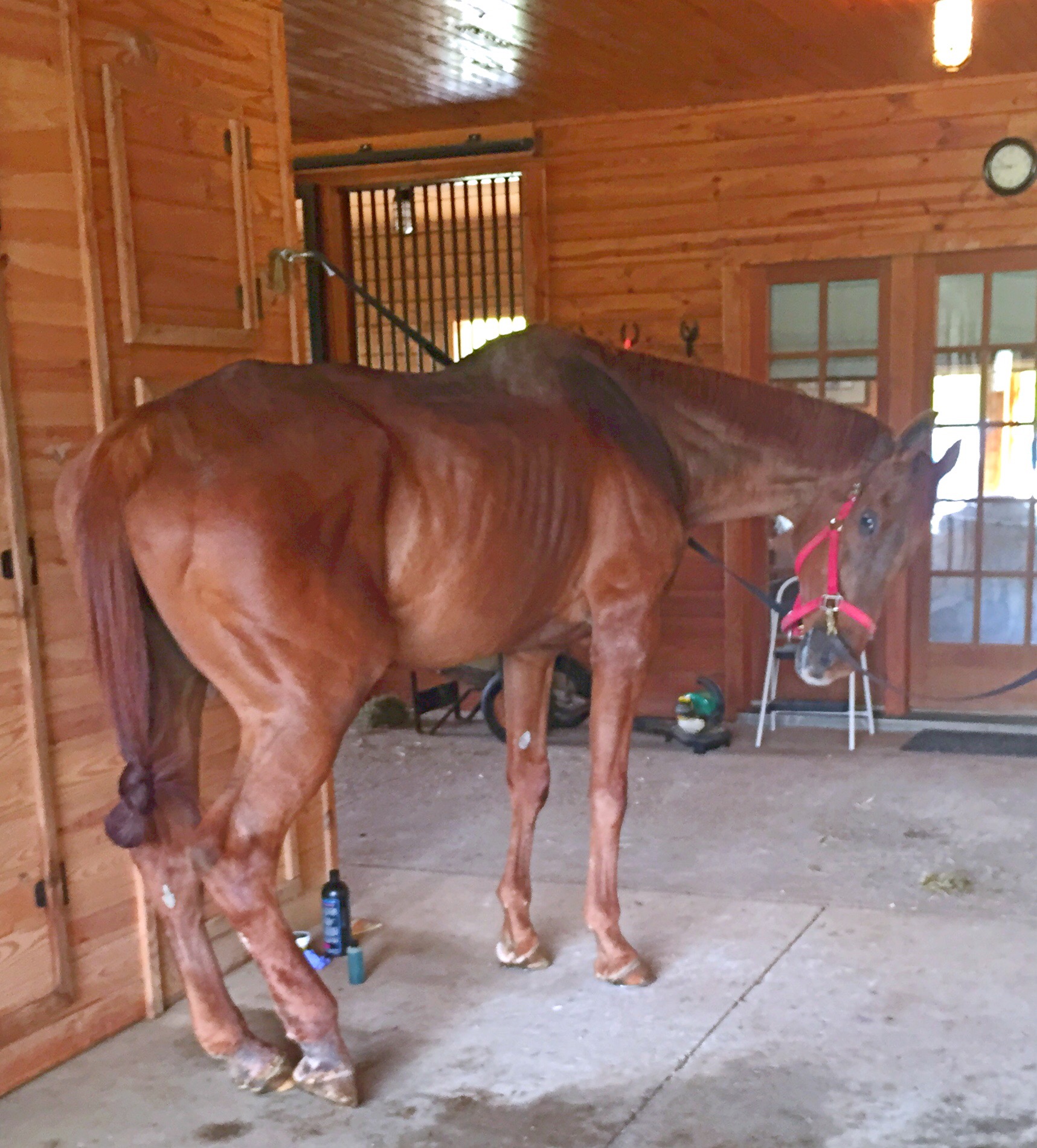
The other day I noticed that Chance’s back fetlock a were slightly swollen and he was visably stiffer then normal. I also noticed a golf ball sized lump in the middle of the his chest. It wasn’t super sensitive and looked like a tick bite reaction, except there was no tick and a tiny barely noticeable scratch. 

I put a Poltace wrap on his back right leg (which was the leg he had previously injured and received stem cell injections in) and gave him some pain medication. I also started him on Baytril and Ulcerguard as a precaution as previously advised by the vet.

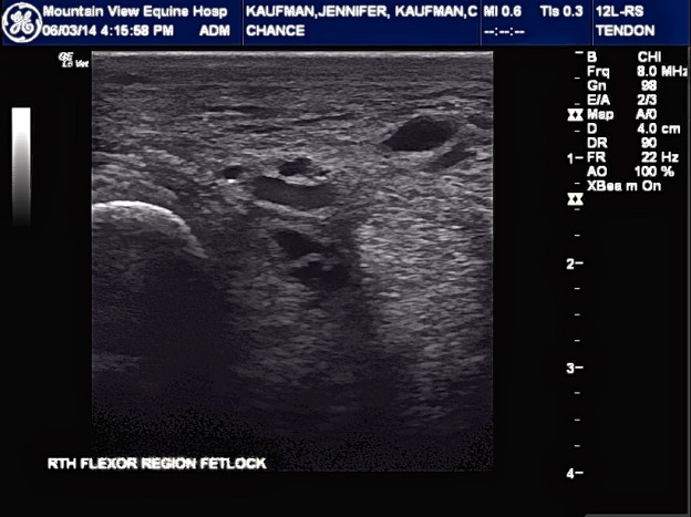
I made an appointment with our vet to come ultrasound his hind right leg and she was to come out in the next two days. I was incredibly anxious to say the least.  

The vet arrived and explained that the lump on Chance’s chest was a hematoma from another horse biting him or from him hitting something. Nothing to worry about, it was just the pooling of fluids to lowest point. 

I then trotted Chance back and forth as the vet watched. After an exam and the ultrasound, the vet explained that she felt that the swelling was due to Chance’s hip pain and the Pastern dermatitis that we have been treating and we’re finally coming off. 

The ultrasound showed a tiny DDFT lesion (vet referred to as a defect that shouldn’t be causing any symptoms). The ultrasound also showed scar tissue that we need to get “stretched out” so that he can gain increased flexibility and work as a protection for Chance’s tendons and legiments. The ultrasound also showed some fluid build up as well. Chance’s Fetlock looks good as do his legiments.

The vet wants Chance to stay on Baytril and Ulcerguard until complete. She also has added a 5 day course of Benadryl and steroids to help with edema of back hind legs. 

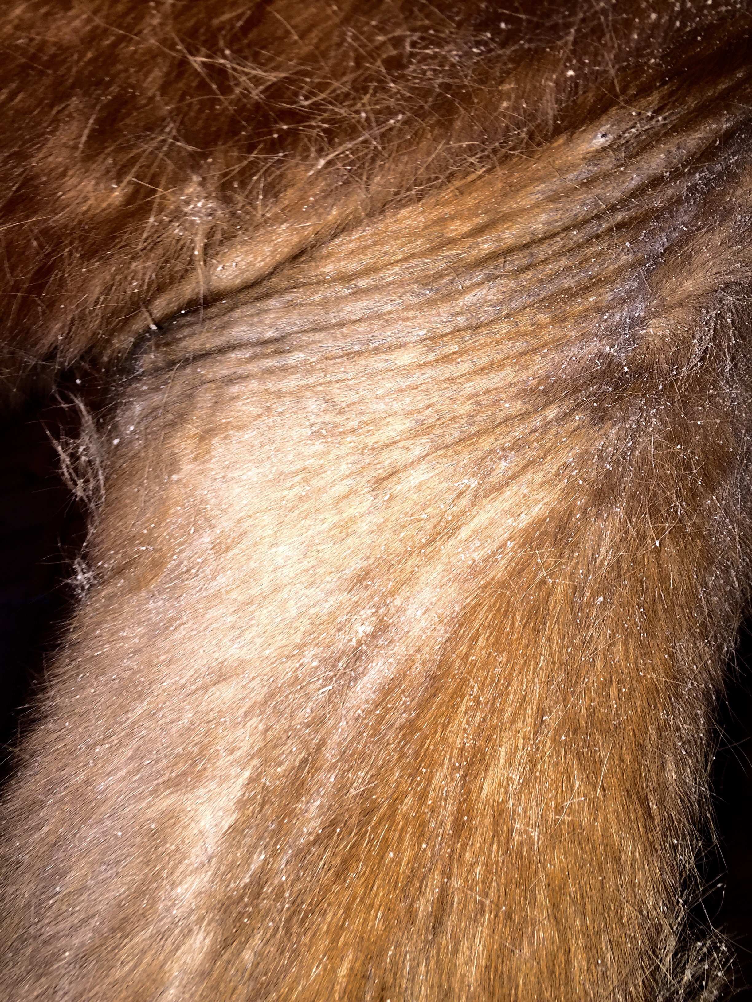
She also provided me with a shampoo that is milder to clean off scratches and apply swat after cleaning. The vet explained that she didn’t understand why people picked the scabs from the scratches because they’re super deep and pulling the scabs off does more harm than good.  

The Vet commented on Chance’s weight gain and how great his skin looks gooded. She wants me to continue working on the scratches and continue doing physical therapy on hills to build up his hind end then get farrier out for back feet. 

All and all I feel good about how Chance is doing and feeling. He is still full of energy, eats like he hasn’t eaten in a week, and his eyes and coat are bright. He is not on daily pain medication and is only given it when he is not feeling great. Aside from a few hiccups, Chance is loving life and being spoiled!

IMG_4332

The BEST Treatment for Scratches 

The other day the vet gave me a way to treat and get rid of my horse’s treatment- resistant, stubborn, and seemingly  IMPOSSIBLE to get rid of scratches or Pastern Dermatitis. 

She suggested mixing Betadine and Vaseline and applying it to the infected area, then wrapping it with Seren wrap and a standing wrap. Leave it on for 12 hours, remove, clean, and redo if necessary for up to 3 days. 

Well, it worked! The scabs just fell off! No more trying to pick off the scabs resulting in the discomfort of my horse or struggling to get him to let me pick at him! 

I have been fighting my horse’s scratches for about 15 years- antibiotics, ointments, MTG, baby oil to soften them, Zinc Oxide, wraps, immune boosters, etc- and nothing has worked until now! 

Trust Your Instincts

Chance being lunged after many years of not being lunged due to a DDFT injury and possible EPM treatments.  He is still stiff and needs to build up the muscle in his hind end thus why he is being lunged.  In addition to his chiropractic adjustments, acupuncture, stretches and massages, and all natural pain medication (MicroLactin), he is also doing physical therapy- walking over poles, walking serpentines, and walking up and down hills.

New Concerns Have Sprung

image

Chance greeting me as I drive up

The days are finally feeling like spring!  The grass is green, the blankets put away, the sun is shining, and the horses are shedding.

Chance is continuing to gain weight, although as I said in the previous post, he still needs to put on a good 50-75 lbs.  As the days continue to get warmer, Chance’s arthritis seems to become more manageable for him; his stride is longer and he runs around (mostly after Lucky) more frequently.

Unfortunately, when the farrier came out about two weeks ago Chance was too stiff to get his back right shoe on. The farrier decided to come back out to try and re-shoe him and, during that time in between, Chance must have tweaked it…AGAIN! 

 While Chance did not have a shoe on his back right I kept it wrapped to provide some protection and also even out all of his hooves.  However, when I arrived I noticed that Chance was significantly twisting his back right leg inward at the walk & it had some swelling.  The swelling was not horrible but it was noticeable. I cold hosed his leg for about 45 minutes while I groomed him & gave him a dose of Equinox (pain medication) and Ulcer Guard.  I put on his back leg wrap to help with reducing the swelling and provide some extra support.  Chance did his neck stretches effortlessly and was baring weight on his back hind. 

But as I was grooming him I noticed, on the left side his chest, he had patches of hair loss and dandruff.  The area did not look red or inflamed, nor did it seem itchy or painful.  So I continued grooming him and decided to put a call into the vet to come and check his leg and the hair loss.

Of course, I turned to Google to try and find out what exactly could be the cause of the patches of hair loss.

image

According to a handful of sources, there are a few possibilities for hair loss- mites or Lice, a vitamin deficiency, rain rot or crud, or even just his natural shedding tendency. A skin scrape would help to confirm what may be the cause. 

 As for the swelling of Chance’s back right leg, I decided to call our previous vet who collected and injected Chance’s DDFT with stem cells to heal the hole in his tendon. We have some stem cells left over and I wanted to see if injecting his leg again would be of any benefit.  I also would like to get an ultrasound recheck to ensure that there is not another injury to his DDFT tendon sheath again.

The twisting of his back hind leg is worrisome as well.  

Everything I have read about EPM states that horses can have a relapse in symptoms after treatment is complete. My concern is that the twisting are due to the neurological symptoms coming back since Chance’s EPM treatment has been finished for a little over two weeks…. 

 Our current vet believes that Chance’s ataxia and twisting is not due to EPM but his cervical spine instead.  Could the twisting be worse due to the swelling of his hind leg?  Or is the swelling and the twisting two separate issues all together?

I pray that gentle hands may guide my feet; I ask for kind commands from voices sweet; At night a stable warm with scented hay, Where, safe from every harm, I’ll sleep til day. -Author Unknown.

Today I met with a “new” vet, Vet7, who was Chance’s very first vet over 15 years ago, when he was vetted at purchase. She came out to do a chiropractic and acupuncture consultation, along with a general check-up. Vet7, while she has a more holistic approach to vetrinary medicinw, she also practices Western medicine.

Vet7 looked at Chance from head to hoof and took a thorough medical history, and ran some tests. She mostly did some balancing and acupressure type diagnostics, and declared that while Chance may have had EPM, she felt that his ataxia was due to his cervical spine….

She explained her reasoning:

1. EPM diagnostics are sub par at best. Even the spinal fluid testing. And, like I previously posted, about 50% of horses in the USA are EPM positive while only 1% are symptomatic.

2. Vet7 did a stretch test with Chance’s neck- to the left, while he compensated by bending at the top instead of the bottom, he showed little flexibility and increased ataxia while doing so. The right side bend was easier. Typically, horses with Cervical issues have issues with the hind-end on the opposite side. Chance has issues with his right leg.

3. Typically horses with EPM respond to treatment. Chance is on his second round of EPM treatment and while his symptoms have had moments where they are less noticeable, there are other moments when his symptoms are prominent; mostly at times of stress. (ie: trailering, new donkey friends, etc).

4. Vet7 put a needle in one of the points in Chance’s tail. He responded immediately showing that he has feeling and strength in his tail, which is something most EPM horses do not exhibit. Typically, an EPM horse will have weakness in his tail, and a times their tail is too weak to lift when they goto the bathroom resulting in having dried manure along their backside.

Vet7 made some adjustments and stuck Chance with a handful of needles. At one point he fell asleep with his head in my arms. She proceeded to inject different points along his cervical spine and hips with B-12.  She taught me some stretches and massage techniques, exercises he and I could do together to increase his hind-end and neck muscles, and gave me some weight gaining instructions.

Chance will have Rice Bran added to his feed beginning with 1 cup for a week and slowly increasing to 2 cups. This will aid in getting his weight up. Apparently, horses are able to tolerate up to 30% fat in their daily diets.

She also suggested adding a Probiotic to increase his Immune system since the gut is the control center. There were other supplements that she felt maybe helpful as well, Cervical Formula, to help with his neck flexibility and overall health.

As for the exercises, in conjunction with turning him out daily, he and I will do stretches to help increase his neck flexion and balance, and I will hand walk him. We will walk on flat land, up and down small hills, and do serpentines. Eventually, increasing to twice a day and adding ground poles and lunging. This will help to develop the muscles, increase his flexion, decrease the ataxia, and aid in his overall health.

acupuncture

Vet7 will come back out in two weeks to see how he is doing…I’m hoping that we have finally found some answers and are closer to a solution.

Lovely Healing

Vet4 came out to Sperryville to do an ultrasound recheck of Chance’s back hind leg.
The stem cell site laceration are healing well. Due to his weight being low, and his difficulty putting weight back on, I asked for a fecal exam to test for any parasites, etc.(The test came back negative.)

I also wanted to do a skin scrape on scratches to try and knock them out for good.

Vet4 said that Chance “looked great” and the laceration on his tendon was almost healed. I receive bute, banamine, and meds for his scratches. I was also provided a name to schedule a consult with a well known nutritionist in the area.

DDFT Lesions

Text from Vet4 after ultra sound #3:

“I can see that he has a deep digital flexor tendon lesion and the medial side of the tendon sheath has improve but lateral side is about the same. The DDFT may the cause of all this in the first place and everything else is secondary. We will re ultrasound in 10 days or so just to confirm my findings. If they are correct, it would help to treat that area.”

Conversation with Vet4 :
So far, Chance has received; shock wave therapy, compression therapy and laser therapy.

Chance has a hole in his tendon. Vet4 believes that this is due to an infection/bowed tendon and severe lameness. The ultrasound, done yesterday, shows no change in size of the tendon hole after the previous two rounds of injections.

“Lymphangitis is a symptom rather than a cause and the cause was never treated.” Vet1 continued to treat it like a disorder rather than a symptom!

The swelling and infection have dissipated, as has the severity of the lameness. Though still lame, he is running around in the pasture.
Pain meds were started again due to increased discomfort and soreness.

Vet4 suggests doing one of the following:

1. Stem cell- which can take about two days if sample drawn has enough stem cells. If not, it could take about 4-6 wks to culture. Once injected he can move home. He is to be hand walked for a few days and then can go out as normal. Vet4 will come out in about a month to do another ultrasound and, depending on the size of the hole, may need to do further injections.

2. Surgery to clean out but NOT repair the tendon. This was not discussed in detail.
Payment plans may be an option. I emailed the office for payment options.

Time to make another decision!

100% Turn Around!

Spoke with Vet4 today. He said Chance has made a “100% turn around”. He trotted him today and Chance was putting full weight on both hind feet! Swelling is disappearing as well!

We spoke about further treatments aside from the Baytril.

I asked about potassium penicillin- He is apprehensive to do potassium penicillin due to horses on antibiotics having DNA changing effects. That it is best to stick to the Baytril and do an ultrasound tomorrow (Friday) to view any changes to the masses. He suggests to have Baytril on hand when Chance leaves to begin immediately if swelling occurs again, which he believes will not be the case after this hospitalization.

I asked about Hydraulic acid: He also is hesitant to inject the SS with the Hydraulic acid due to it’s effects on certain bacterial strains- often allowing the bacteria to hide from the antibiotics. He does agree that another round of injections would probably be helpful and will know more after the next ultrasound.

When asked whether scratches can lead to Lymphangitis, thus leading to the infected SS, he said it is hard to tell but certainly possible.

Chance is currently receiving laser therapy and cold compression therapy along with Baytril, pain meds, and supplements.

Vet4 believes that Chance should be able to leave within a week to two weeks depending on progress!

Dr. Fortier’s “Lameness Originating from Tendon Sheaths”

Lameness originating from tendon sheathes.
Lisa A. Fortier, DVM, PhD, Diplomate ACVS Cornell University, Ithaca, New York, USA
Laf4@cornell.edu

(Below is cut and pasted from Dr. Fortier’s “Lameness Originating from Tendon Sheaths”)

The most commonly affected tendon sheathes associated with lameness is horses are the digital, carpal, and tarsal sheaths. In general, the diagnoses of lameness originating from tendon sheathes is increasing with awareness and with the more common use of MRI. Clinical signs associated with tendon sheath lameness are variable with respect to degree of lameness and extent of synovial distension. In general, the lameness will worsen with flexion and with work, but the degree of lameness is not directly associated with prognosis for return to athletic performance.

Lameness associated with the digital sheath

Lameness can be localized to the digital sheath with a low 4-point block or intrathecal anesthesia. If sepsis is suspected, a clean (non blood-contaminated) synovial fluid sample can be obtained at the base of the sesamoid bones and axial to the palmar digital neurovascular bundle.

Simple annular ligament constriction, without involvement of the superficial (SDFT) or deep digital flexor tendons (DDFT) is a common cause of lameness associated with the digital sheath. When viewed from the side, the palmar/plantar profile of the digital sheath will have a “notched” or “cut-in” appearance at the fetlock joint. The integrity of the annular ligament (thickness and structure) should be evaluated using ultrasonography to be sure there are no other structures involved such as the SDFT or DDFT. Horses can be treated with intrathecal hyaluronic acid with variable success prior to surgical intervention. If the annular ligament is the sole structure involved and there are no adhesions within the tendon sheath, then a closed or semi-open annular ligament transection could be performed rather than a tenoscopic transection. If ultrasound examination reveals adhesions or synovial masses within the sheath, then tenoscopic exploration and removal of the masses/adhesions is warranted.
Performing surgical maneuvers or exploratory surgery under tenoscopic guidance has distinct advantages as compared to open approaches. Tenoscopy allows for more complete examination of the entire tendon and tendon sheath, resulting in a more accurate diagnosis than can be provided by ultrasonography and the surgeon has an opportunity for removal of pathologic tissues such as synovial proliferative masses, hyperplasic synovial tissue, and adhesions. Additionally, the use of tenoscopic portals instead of an open approach reduces potential iatrogenic damage to neighboring structures and decreases the incidence of postoperative synovial fistulation. The biggest take-home message of these notes should be that the presence or extent of synovial masses/adhesions is not directly correlated with prognosis and many horses return to full athletic performance after tenoscopic surgery and removal of adhesions/masses and annular ligament transection.

Longitudinal tears in deep flexor tendon are increasing commonly diagnosed. Like adhesions and masses, tears are frequently worse on tenoscopic exam than on ultrasound. In these cases, the tendon tear appears to be the primary cause of the tenosynovitis and the annular ligament constriction is likely secondary. Currently, debriding the tendon edges is the only treatment, but some tendon repair technique seems warranted. Ian Wright described a combined approach to repair these tears, but has subsequently discontinued this practice and simple debridement of the granulation tissue between tendon edges is recommended.

Waiting Games

We began Baytril on 8/16.  The next day Chance came in from pasture with NO fever, NO trouble walking, but also NO appetite- eating a little grain, hay stretcher, peppermints and the swelling worse.

Chance got Compounded Baytril- 2 scoops with feed previous pm. Vet3 advised us to give another dose of Baytril but Chance won’t eat (most likely due to the taste of the Baytril in feed previous night. But Vet3 believes it is due to his pain). So, we gave another dose of Banamine/10 cc (am and pm) and Tridex- 1 packet. Iced 2x/kept in/ wrapped both hind legs with boots.  And the waiting game begins!

Research, Research, Research

Tendon Injury Handbook

After I left the barn, I drove home and went straight to my computer.

What was happening?  What are the masses? Scar tissue?  Nothing was able to be extracted out of them…How can I get rid of them in order to see behind them?

Again, I stayed up until the sun came out the next morning.  I already had two binders full of research and now I had a third.

Research made me believe that C has an infection in the Synovial Tendon Sheath that was being masked by the masses on the outer lining of the SS. The masses could be scar tissue from his MANY past Lymphangitis flare-ups. Perhaps, his immune system was not able to fight last attack and the infection settled in the SS and was walled off.  Thus his CBC & WBC were normal and no fluid was extracted from SS masses due to the large size of the scar tissue.
C has a major hx with his RH and “flare-ups” and lameness. I never realized this until I took the time to study his past records from the first 5 years I owned him.

Symptoms are similar to an infection- what if we proceeded as if it were?
Lack of a positive culture does NOT mean that there is not an infection in the sheath!

Current Symptoms:
1. Swelling decreases after being active
2. Fails to extend fetlock
3. Lame- exasperated by flexion
4. Positioning for fetlock flexion

Septic Synovitis: Cartilage degradation ischemia, Fibrin deposition lead to lameness to pannus form and adhesive form

Entrobacteriacaea
Strep
Staph
Most common is Staph

Treatment: 

Systematic Procaine Penicillin 22000 iU/kg or Sodium Benzyl Penicillin & Gentamicin 6.6 mg/kg for 2-9 days

Then change to oral potentiated sulfonamides 5mg.kg Trimethoprim and 25 mg/kg of Sulphadiazine

Other potassium penicillin w/ Amikacin Cectiofur or Enrofloxacin

IV antibiotics for 7-10 days switch to oral for 2 weeks

Regional limb profusion or placement of impregnated Polymethyylmethacralate or PMMA

I immediately called Vet4 and told him my theory.  He said that it was possible and that we should begin treatment asap.  He was still out of town so I called Vet3 to order Baytril. Vet3 felt my theory was legit and immediately ordered the antibiotic!

“Ask him what he wants…you will know what to do from there.”

When I arrived at the farm I was greeted by those familiar big brown eyes and a whinny.

I brought him out of his stall and gave him a bath.  He has always loved to be groomed and bathed, even if he is apprehensive to walk into the wash stall. After his bath, we went outside for him to graze and get some sunshine.  When we walked inside the barn, I stopped him and looked into his eyes and asked him, “do you want to keep trying to get better or are you ready to give up?”  He just looked at me.  A lump immediately formed in my throat.  He nuzzled me and rubbed against me.  I said again, “Do you want to keep fighting?”  He shook his head up and down! I swear to you!  This actually happened!

The decision was made, we would keep on fighting as long as we were able to keep him comfortable.  There was nothing I wanted more than to bring him home with me, let him live out the last days of his life pain-free, and with me by his side.

This was his turn.  He had always done what I asked of him- lessons, moving stables and even cities, and he was patient while I was in school- and it was his turn for me to make it about him.  For Chance to get every ounce of my time and for me to fight for him!

IMG_1795

Road blocks


IMG_1892


The first round of injections provided Chance with some relief, in his ability to move around and the swelling went down a bit, but that only lasted about a week.  At about day 8, Chance was swollen again and 3 legged lame.  Thankfully, a family friend of the farms owner called me.  She explained that the farrier was out shoeing one of the horses and saw Chance’s leg, and when he arrived to shoe her horses, he expressed his concern.  I immediately contacted Vet4 an he was out the following day.

Vet4 injected the masses again as a temporary fix, until we could make some decisions.  The ultrasound showed that the masses were the exact same as they were in the first ultrasound- they hadn’t increased or decreased in size.

Later that evening, Vet4 and I had a lengthy conversation about where to go from here.

We discussed the options again, at length.  We could do an MRI to gain more insight into what is happening with that leg, go in with an Arthroscope and clean it out, or look into Stem Cell Therapy.

Well, I wasn’t comfortable putting Chance under anesthesia…he was too old and too frail.  Plus, he could break a leg or a hip going down.  So, that ruled out the MRI (unless I could find a standing one) and the surgery.  The Stem Cells would run about $3000.00, plus he would need to goto the hospital to have the procedure done.

I took the night to think it over, and stayed up until sunrise reading as much as I could on leg issues, the different options vet4 and I had discussed, and other potential causes.

That next morning, I received a call that Chance was worse.  Vet4 was out of town due to an emergency, so I called Vet3.  She got out to the farm immediately.

Vet3 gave Chance Surpass topical to put on the leg, Banamine, Ulcer Guard, and continued with the Prevacox to keep him comfortable.

I asked her what she thought about the options- she felt, as I did, the surgery wasn’t a good idea and that an MRI should only be done without sedation.

I called Vet4 and we spoke about the current situation.  What else is going on? He suggested changing the course and trying different diagnostics.  He explained that TSMs (Tendon Sheath Masses) can cause swelling and pain, but they are usually relieved by the injections.  The ultrasounds showed that his suspensory tendon and ligaments looked good.  Could this be an infection? Soft tissue damage? A bone issue?

I asked him if he felt moving forward with more tests was a bad thing…was I being cruel keeping Chance alive like this?  Something that had been weighing on me from the start.  And what Vet4 said, empowered me to continue down the path I initially felt in my gut to be the right decision.  He said, “I am not the kind of person to ever give up on someone or something.” I asked if we were able to manage his pain adequately and make sure he was comfortable and he said, yes.  He advised me to “make a decision based on the horse” and “not to listen to the opinions of everyone else”.

The next day, I cleared my schedule, and headed to the farm.


Coming Home to Love & Peace

Chance was able to come home a week later.  Sam & John went to pick him up at the hospital. However, upon arriving, they soon found out that Chance did not want to get on the trailer.  Sam later told me that one of my sweatshirts was in the truck, so she brought it out and let him smell it- he finally loaded.

I got his stall ready- tons of fresh shavings, hay, a new water bucket…

The moment he got off of the trailer he was shaking!  I gave him a warm bath and let him out in a small, flat paddock so he could be in the sunshine.

This is what happened when I let him out!  He did something he had not been capable of doing for months and months, if not longer.

Answers

Vet4 came out immediately.  I was beyond grateful!

He did an ultrasound of the back right leg and called me.  He found that Chance has Chronic Cellulitis and that there was Vascular constriction, and masses on the tendon sheath between the superficial and deep tendon sheaths.  The Doppler showed good blood flow and a thickening of the synovial lining. Hoof testers- Negative

We spoke about my opinions- MRI, Arthroscopic surgery, Regional Diffusion, Cold Compression Therapy, Nerve Block Injections, Steroid Injections

We decided  to try the Steroid Injections into the 3 Synovial masses to hopefully reduce the size and thickening.  Thus allowing us to see behind the masses to see what is actually happening.

Injections were into the Proximal Digital Flexor Tendon Sheath with 6mg Betamethasone and d100mg of Amikacon. Leg was covered with SSD and DMSO and bandaged.

Once injected, cold compression therapy for about 5 days twice a day and stall rest. Banamine daily.