Category Archives: Symptoms

I Loathe Ticks! 

Your horse comes in from being outside and is barely able to move.  His legs are swollen, he has a fever, is sensitive to the touch, and has a loss of appetite.  He has chills- intermittently shaking.  He wont touch his hay, his eyes are dull, and he looks depressed and tired.  You call the vet and they run hundreds of dollars worth of tests- CBC, x-ray his legs to ensure there is no fracture; they diagnose him with Lymphingitis.  You begin a course of antibiotics.  You cold hose.  You give him Banamine.  Your wrap his legs while he is on stall rest. A week later, the swelling has subsided, his fever has dissipated, and his appetite is back.

You get a text saying that your horse “ran away” when he had been let out earlier that day.  But when you get to the barn, you notice when he turns he looks like his hind end is falling out from under him..remember when you were little and someone would kick into the back of your knees and your legs would buckle?  That is what it looks like.  So you watch him.  You are holding your breath, hoping he is just weak from stall rest.  You decide, based on the vet’s recommendation, to let him stay outside for the evening.  You take extra measures- leaving his stall open, with the light on, wrapping his legs, etc- and go home.  Every time your mind goes to “what if..”, you reassure yourself that your horse is going to be okay and that you’re following the vet’s advice and after all, your horse had been running around earlier that day.

The next morning your horse comes inside and it takes him an hour to walk from the paddock to his stall.  All four legs are swollen.  He has a fever (101.5).  He is covered in sweat.  He won’t touch his food.  He has scrapes all over his body and looks like he fell.  You call the vet- again- and they come out to look at him.  They note his back sensitivity, his fever, the swelling at his joints (especially the front).  They note that his Lymphingitis seems to have come back.  The vet draws blood to check for Lyme.  They start him on SMZs and Prevacox.  You once again wrap his legs, ice his joints, give him a sponge bath with alcohol and cool water to bring down his fever.  You brush him, change his water, put extra fans directed at his stall.  You put down extra shavings.  And you watch him.

A few days go by and you get a call saying that your horse has tested positive for Lyme…and while your heart sinks, you are also relieved that there is an explanation for your horse’s recent symptoms. You plan to begin antibiotics and pretty much not breathe for the next 30+ days while your horse is pumped with antibiotics.  You pray that he doesn’t colic.  You pray that you have caught Lymes in time.  You pray that the damage is reversible.  You research everything you can on the disease.  And you sit and wait….

Below are resources on Lyme Disease in horses- treatments, symptoms, the course of the disease, and the prognosis.

epm-diagram

Lyme Disease in Horses | TheHorse.com

Lyme Disease, testing and treatment considerations | Best Horse Practices

Microsoft Word – Lyme Multiplex testing for horses at Cornell_2-12-14 –

Lyme_Disease_Multiplex_Testing_for_Horses.pdf

Fall Fever

Today Chance had swelling of his back right fetlock.  He had a fever around 104 and didn’t eat his feed.  His eyes were dull and he was lethargic.  He wasn’t limping but was walking slower than normal (he usually runs to the paddock or back to the barn).  I decided, due to the Lymphingitis flare up on his back right leg, I would give him a shot of 5 mls (or 5 cc) of Banamine and wrap his leg.  Once the medication set in, I would bring him in to give him a bath (it was 80 degrees today).  So, that is what I did.  By the time he was back at the barn he was covered in sweat.  I cold hosed him and drenched the wrap in cool water and let him roam around the barn.

Thankfully, the vet was able to meet me at her veterinary practice so that I could pick up Baytril and more Banamine.  Since Chance just had Lyme Disease (and had finished his medication less than a week ago), we are not 100% if this is a Lyme reaction or something else.  The plan is to administer 25 cc of Baytril either orally, in his feed, or via IV for 6 days and Banamine 10 mls (or a 1000 lbs) twice a day for 3 days. The vet suggested that I do 5 cc of Banamine if his fever remains between 101-103 degrees and 10 cc if his fever is 103 degrees or above.   During this time I will begin Prevacox- one 1/4 of a tablet once a day.  After 3 days, I will discontinue the Banamine and continue the Prevacox.  If his fevers are not down in two days, I will continue the Baytril but start the doxycycline as it maybe a Lyme disease symptom.

While researching Lyme Disease, I found that many people do two+ months of doxycycline instead of 30 days to ensure the disease has been erraticated completely.  However, since Chance had shown such improvement after 30 days, I decided to not do another month.  Maybe I should have…

However, Chance had similar symptoms when we found a small laceration in the DDFT tendon of his back left hind- swelling, Lymphingitis, fever, lethargy, no appetite, etc.  If he does have an issue with his tendon I will most likely do another round of Stem Cell treatments which proved to be helpful last time.  Thankfully I stored his stem cells in a Stem Cell Bank (via Vet-Stem) and can easily have them shipped.


Guttural pouches: The hidden dangers in horses

An interesting article I came across about Guttural pouches in horses.  Must read for those who work with or around horses.


https://apple.news/AisI_D1YlPNetjlUilRFbaA

EPM Tilter. What Do The Numbers Mean?

During my horse’s recent Lymphingitis flare-up, the vet advised that we run labs to test for Lyme and EPM due to his presenting symptoms (hind weakness, twisting his back leg at the walk/walking sideways I refer to it as- “Chance’s swagger”).  As I noted previously, Chance’s Lyme test revealed that he was at the beginning stages of an acute infection…yay for the labs at Cornell University for their amazing ability to give you more than a positive or negative!

A little history before getting to the EPM Tilter results.

About 2ish years ago, Chance was diagnosed with EPM (and one of the reasons opossums and I are not friends since they host the disease as do a few other culprits).  Chance immediately began EPM treatment- he received Protazil in his feed for one month. After hours of research I chose Protazil, although extremely expensive (if you order from http://www.drfosterandsmith.com they sometimes have promotions where you receive store credit for every $100.00 you spend…they did when I ordered and I got a “free” dog bed that my dogs adore), due to the decreased likelihood of Chance experiencing a “Treatment Crisis” (worsening of symptoms) and the ease of administration (other brands require the drug being administered 1 hour before eating or an hour after and so on).  Typically, EPM treatment is done for 30 days and, depending on the residual symptoms, some may require subsequent treatments.  While Chance’s symptoms improved, I wanted to ensure that we annihilated the disease and did another round of treatment but this time with Marquis.  At the end of two months, Chance’s ataxia was gone!

Fast forward to September 2016…Chance, just having a Lymphingitis flare-up, has been tested for Lyme and EPM. Lyme came back positive.  And….so did the EPM test..well, kind of.  Wonderful.  (See why I loathe opossums?)

Chance’s EPM test #2 on 8/30/16 (the 1st one was 2ish years ago) showed the following:

“Combined SAG 2,3,4 Tilter on serum= 1:2000”

So, what does this mean?

The test revealed that Chance had “positive, specific antibodies” detected in the blood work.  This means that he had EXPOSURE to S. Neurona, a causative agent of EPM.   Serum tilters range from <1:250 (negative) to >1:4000 (high positive).  S. Neurona (SarcoFluor) is one of two protozoa found in EPM infected horses, the other protazoa is N. Hughesil (NeoFluor). S. Neurona is most frequently seen, whereas N. Hughesil is not as common.

The vet ran another EPM test to confirm the findings in the 8/30/16 test.  The results showed that Chance had “Combined SAG 2,3,4 Tilter on serum= 1:1000.  Again, Chance showed EPM protozoa in the positive-ish range.

I initially had not seen the results but was told by the vet that he was EPM negative.  So when I asked for the test results to be emailed to me and saw the numbers I sort of freaked out…I emailed the vet to ask for clarification.  She explained,

“The EPM test shows that he was exposed to the organism in the first test we did which is why we did a follow-up test. Since his exposure level dropped from 1:2000 to 1:1000 this shows that he does not have the disease. There is no good one time test for EPM once they are exposed which is why we had to do the repeat to compare the two.”

While this explanation offered me comfort, I was confused…why does he have any protozoa in his blood if he doesn’t have EPM?

I spoke to another vet and she explained it in a bit more detail…I am hoping I am summarizing what she said correctly..

When a horse tests positive for EPM they either have an active disease or they may not.  However, when the test does from 1:2000 down to 1:1000 this typically means that the horse’s immune system is working correctly to fight the disease off- active or not.  EPM testing typically provides you with a % of the chance your horse has an active EPM infection, or at least if you send it to Cornell University.  For instance, lets say a horse gets the results back and it shows that they are “positive” or have been exposed to S. Neurona (one of the two EPM protozoa)…their results are  1:647.  This means that, after doing a bunch of adding and multiplying that this vet kindly did for me, the horse has a 60-70% chance of having ACTIVE EPM.  Meaning, he most likely would be symptomatic (ie: behavioral changes, ataxia, weight loss, difficulty eating, changes in soundness, and a bunch of other neurological symptoms).

My hunch is that Chance’s immune system was boosted because I started him on Transfer Factor (amazing stuff… more information can be found in some of my older posts) again as soon as his results came back positive for Lyme.

Here are the 3 EPM tilters that were run on Chance (oldest to most recent) along with his Lyme test results:

epm-2014

epm-test-results-8-31-16

EPM results 9-8-16 copy.jpg

lyme-titer-8-30-16

I have a limp!

Resources on how to diagnose, treat, prevent, and handle lameness in horses

Your Horse Has a Swollen Leg – Why and What To Do | EquiMed – Horse Health Matters

All About the Fetlock

Fetlock Lameness – It’s importance… | The Horse Magazine – Australia’s Leading Equestrian Magazine

Causes of Equine Lameness | EquiMed – Horse Health Matters


Common Causes of Lameness in the Fetlock

fetlock lame


Equine Podiatry

Medical History


  1. DDFT Lesion on right hind
  2. Cervical Spine Arthritis
  3. Hip discomfort due to past fall

Past Treatments Tried


  1. Stem Cell Injections: Healed the DDFT lesion in right hind until recently the lesion began to reappear
  2. Ozone Therapy: Assists in the healing of tissues
  3. Shock Wave Therapy: Assists in the healing of tissue
  4. Chriopractic adjustments
  5. Acupuncture
  6. Supplements

Initial Consultation


Chance showed decreased movement in his right hip and a audible cracking noise at the suspensory joint.  He has edema of both hind fetlocks, Pastern, and Pastern Dermatitis.  Chance was unshawed on both hinds due to his inability to stand for long periods of time and his decreased mobility.  However, his front adorned clips.

Due to the length of Chance’s front toes and the height of his heels he was unable to evenly distribute his weight (60/40) to his front and hind ends.  This would most likely cause increased tension on the DDFT tendons and corresponding ligaments resulting in an increased likelihood of tendon and ligament related injuries.  The uneven distribution of weight could also inhibit the horse’s range of motion through his hips resulting in his body compensating for this injury and causing ataxia (balance issues), pain, arthritic changes, and cervical spine misalignment.

By shortening the toe of both front feet, the heel will rise allowing a more even distribution of his weight.


Front


IMG_3193

IMG_3196


Final Product: Front


Trimmed feet to corrected to the following specifications:

Foot   Beginning Angle & Toe       Corrected Angles & Toe   Total P.C.

L/F    47 Degrees at  3 7/8 inches   53 Degrees at 3 inches         6 Degrees

R/F    45 Degrees at 3 3/4 inches    54 Degrees at 3 inches         9 Degrees


Hind


20160604_160438


Final Product: Hind


20160609_153622

 Return visit to trim and shoe Chance’s hind feet with #2 OBRHB Wedge shoes.Trimmed hind feet and corrected to the following specifications:

Foot   Beginning Angle & Toe       Corrected Angles & Toe   Total P.C.

L/H   48 Degrees at 3 7/8 inches    54 Degrees at 3 1/4 inches    6 Degrees

R/H  46 Degrees at 4 1/4 inches     55 Degrees at 3 1/4 inches     9 Degrees

Note: Chance needed to be sedated by veterinarian to complete the trim and shoe his hind feet due to preexisting hip and DDFT issues.

Ozone Therapy


OZONE THERAPY – HISTORY, PHYSIOLOGY, INDICATIONS, RESULTS


 Judith M. Shoemaker, DVM 305 Nottingham Road Nottingham, PA 19362

717-529-0526 Fax 717-529-0776

http://www.judithshoemaker.com

Ozone therapy has been utilized and heavily studied for more than a century. Its effects are proven, consistent, safe and without side effects. Why is it not more universal in its use? Many of you have come with some trepidation about infusing a gas into a vessel because you are concerned about emboli, or have some dreadful fear about ozone’s toxicity since we frequently hear about the unhealthy ozone levels in the atmosphere. These fears do not apply to properly administered medical ozone, and the potential benefits of ozone therapy are profound and without associated detrimental effects.

Oxygen, in its several forms, cycles through the atmosphere and life processes just as water does. Ozone is produced in the upper atmosphere when UV light strikes the oxygen rising from plants, plankton, and algae in our forests and seas. It then falls back through the atmosphere, as it is heavier than air, combining with pollutants and water, cleaning the air and forming peroxides that benefit plants. Ultraviolet light breaking down pollutants and nitrous oxides also can produce ozone at the ground level, which is the eye and lung irritant in smog.

Medical ozone, used to disinfect and treat disease, has been around for over 150 years. Used to treat infections, wounds, and multiple diseases, ozone’s effectiveness has been well documented. Ozone has been used to disinfect drinking water since before the turn of the last century. A text on medical ozone therapy was published by Dr. Charles J. Kenworth in 1885! The best technology for producing ozone gas was designed and built by Nikola Tesla in the 1920’s. Heads of leading medical institutions in the U.S. contributed to a 1929 book “Ozone and Its Therapeutic Actions” describing the treatment of 114 diseases using ozone.

In 1933, the AMA began its systematic suppression of all modalities of treatment that did not complement its liaison with the emerging pharmacologic and diagnostic industries. Ozone therapy, along with many other useful therapies, were methodically eliminated from the educational process and exposure to the public in the U.S.

Less suppression has occurred in Europe and other countries, especially in Russia. Today in Germany, and other countries, ozone therapy is commonplace. Over 7000 doctors in Germany use it daily. In fact, in Germany, ozone generators are in ambulances for treatment of stroke victims. The incidence of permanent paralysis in these patients is much less than that in similar patients where ozone is not used.

Ozone generators are relatively simple and inexpensive. The equipment used to handle ozone is readily available but needs to be relatively non-reactive. Glass, Teflon, Kynar, silicon, and gold are completely non-reactive. Equipment made of other substances can contaminate the ozone or just deteriorate rapidly using up the ozone and becoming nonfunctional.

 Generators use several technologies to produce ozone

  • UV lamp – makes small amounts of ozone and is unreliable in making accurate concentrations. They burn out easily.
  • Corona discharge – dual dielectric sealed systems produce ozone but also lots of heat which is both destructive to ozone and to the machine.
  • Cold plasma generators – which produce ozone using low level current passed in 2 tubes of a noble gas between which an electrostatic plasma field forms that ionizes the oxygen.Ozone concentration is measured in u/ml or gms/L of oxygen, 5% or 70 u/ml is usually the maximum concentration used in clinical medical applications. High concentrations will damage red cells and inhibit growth of healthy cells.Dosage and frequency protocols vary widely. Initial high dose treatments may “jumpstart” the immune system followed by lower doses. Those who are fearful have been “starting slow and going low” with dosage and still have good results. Concentration must be carefully controlled with accurate flow rates, requiring pediatric regulators for the needed slow flow rates to produce high concentrations. Therefore, home made machines and lesser quality nonmedical devices are not appropriate.

    Ozone poteniates free radical scavenging substances and systems in the body, inducing the production of superoxide dismutase, catalase, and glutathione peroxidase. If ozone administration causes any respiratory irritation from out-gassing through the lungs, a bolus dose of 1 to 5 grams of vitamin C can be given and will eliminate any coughing instantly.

    Oxygen/ozone mixtures cannot cause emboli when injected at reasonable rates as they dissolve and diffuse very quickly in body fluids, unlike air (predominantly nitrogen) which is what forms emboli and causes the bends or decompression disease.

    The physiologic actions of ozone are many, the simplest of which is to provide sufficient oxygen to allow complete oxidation of sugars and other fuels to produce sufficient and efficient energy and to “burn clean” to CO2, water, and inert end products. If not enough oxygen is available, then incomplete oxidation occurs, producing carbon monoxide, lactic acid, and partially oxidized toxins that inhibit further oxygen metabolism and “clog the system”, tying up hemoglobin, water, and the mechanisms for function and elimination.

    Administration can be through any route with modifications:

  • Direct IV infusion – Ozone slowly administered into a major vessel.
  • Major auto-hemotherapy – Anticoagulated blood is mixed with ozone and is infused into a vessel.
  • Rectal/vaginal insufflation – Humidified ozone is administered by catheter.
  • Minor autohemotherapy – Blood mixed with ozone is injected intramuscularly.
  • Limb or body bagging – Body or parts are bathed in humidified ozone.
  • Ozonated water – Dissolves easily in water to be used topically or consumed.
  • Ozone in Saline or LRS – Can be used topically or given IV or SQ.
  • Intra-articular administration – For joint healing and prolotherapy.
  • Prolo/Sclerotherapy – Very good, less painful than other agents.
  • Acupuncture – With ozone, more effective than B12.
  • Ozonated olive oil – Ozone is bubbled through oil until the oil is thickened. This will produce ozonides that are not irritating and thus is applied topically even to eyes.
  • Inhalation – Ozone that has been bubbled through olive oil and humidified will not irritate respiratory epithelium.
  • Subconjunctival injection – For ulcers and keratitis sicca.
  • Gingival and tooth apex injection – Can eliminate infection.
  • Urinary bladder insufflation – For chronic inflammation.
  • Disc protrusions – Prolotherapy, which can be injected at interspinous space and around facets, stabilize joints and accelerate healing.
  • Auricular – Can be direct, humidified, or bagged with a homemade device made from IV bags and tubing (á la Margo Roman).As an anticancer, antiviral, antibacterial, and ant fungal agent, ozone is unsurpassed, especially since there are no appreciable side effects.Oxygen deficit is key to the development and progression of all disease processes. Dr. Otto Warburg’s work, confirmed by others, shows that this deficit and subsequent toxin buildup is the fundamental cause of all degenerative disease, especially cancers.

    Antioxidants help the body to protect itself from excessive oxidative damage caused by multiple free radicals, many of which are inactivated by ozone. The support of free radical scavenging systems is important but only oxygen can improve the deficit that makes cells vulnerable to oxidative damage in the first place. Long-term ozone therapy can be augmented by supplementation with antioxidants, but normally they should not be administered within 4 to 12 hours of ozone therapies.

    Ozone produces the same effects as exercise, which produces significantly more free radical oxygen than can be administered in any ozone treatment. Ozone equals ”exercise in a syringe” without doing joint damage.

    Ozone potentiates more complete oxidation, helps to maintain more normal body temperature and increases the effects of most hormones, vitamins, herbs, homeopathics, and drugs. Concurrent ozone administration reduces the amount of chemotherapeutic drugs needed to achieve effect by 1⁄4 to 3⁄4. It complements chelation therapies and frequently improves the affect and sense of well being in patients.

    Continued therapy will allow Herring’s Law to manifest “Healing from inside to outside, top to bottom, front to rear, and in reverse chronological order of the insults to the body.” Healing crises, however, may occur. Ozone therapy facilitates the rapid resolution of these crises.

    2005 Judith M. Shoemaker, DVM

Treating Cellulitis in horses

A common and potentially hazardous infection that can be helped with combining antibiotics, cold hosing, and exercise.

Liz Goldsmith's avatarEQUINE Ink

Freedom&#039;s left knee was very swollen. You can see the scrape on the outside of the knee.

On Monday night I showed up at the barn around 7:00 p.m. to ride once the day cooled off. To my dismay I found that Freedom’s knee was hugely swollen. I had wrapped his legs because he’d felt a bit stocked up the night before and because the swelling had no where to go except the knee, the effect was horrifying. Visualize a grapefruit.

Almost immediately I saw the cause: a scrape less than an inch long. Before I called the vet I scrubbed the wound to make sure it wasn’t a puncture wound (didn’t look like it to me, but a puncture would where a joint is involved can be very serious). Normally I would also shave the area around the cut but I had sent my clipper blades out to be sharpened!  My guess was cellulitis but since it involved a joint I wanted to makes sure I…

View original post 499 more words

If You Experience Worsening Depression…

Chance began another round of Excede to get his scratches under control- it is a never ending battle.  A while back, I had a skin scrape of Chance’s scratches due to their chronic nature.  The scrape results showed a number of bacteria, all commonly seen with this type of infection, that were resistant to most antibiotics.  Thus why we decided to try Excede.

Administering Excede is pretty straight forward- 1 shot IM every 4 days for about a month.  Easy enough….or so I thought.  The first shot was administered by the vet when I was not present.  The second shot the vet also administered while I was there.  Thirty minutes after the shot was given to Chance I noticed he seemed off but not in his “normal” post-acupuncture relaxed state. He suddenly became lethargic, he wouldn’t eat his dinner, and the gut noises became almost nonexistent. I commented to the vet my concerns and she came over and reexamined him.  Sure enough something was wrong.  She proceeded to administer 10cc of Banamine (just in case it was colic) and told me to walk him around outside for about 20-30 minutes.  Then see if he would eat 2 cups of feed only.  We walked and Chance began to act like his normal happy go lucky self.  Once inside he started to eat!

Part of me felt that his reaction was a fluke.  However, the third dose proved me wrong.  Four days later, Chance received his shot and went outside to enjoy the first beautiful, warm day.  I sat in the field watching him.  He was sluggish, lethargic, stiff..he looked 10 years older and barely moved from one spot under a tree.  He wasn’t eating grass nor did he run around and play- he didn’t even run up to me like he normally would. I decided to bring him inside and give him a warm bath since it was in the high 70’s.  He was non responsive to his bath- no playing with the hose or even accepting peppermints.  I placed a cooler on him to ensure he stayed warm until he was out in the sunshine.  I figured after a bath he would perk up- again, I was wrong.  At dinner time I went to bring him in and typically I will open up the gait and he will canter into his stall- he slowly walked instead. He wouldn’t eat his feed (he normally whinnies and makes a fuss until he gets his feed and devours it) or his hay…I stayed and watched him for a while and he just slept.  I spoke to John, the guy who helps me with Chance and Lucky, and he confirmed that Chance hadn’t been finishing his feed and wasn’t running when he brought him in for dinner.

My concerns grew and I decided to do some research on Excede. That strange thing is I usually do extensive research before changing or administering anything with my animals.  But, for some reason I did not do so this time and I wish I had.

According to a number of reputable websites, Excede can cause significant and dangerous side-effects such as; diarrhea, severe acid reflux, blood coming from mouth, loss of appetite, lethargy, muscle and gait stiffness, and more.

The most troubling of everything that I read wasn’t what was posted on the Pfizer (the manufacturer) website but from the countless statements given by horse owners and the studies done by outside companies.

According to drugs.com, “in the PK study, several horses developed clinical signs consistent with foot pain (stiff in the front limbs when turned in tight circles, and increased pulses and heat to the front feet). One horse in the NAXCEL group and one horse in the 6.0 mg/lb (2X) EXCEDE group were euthanized due to laminitis. Clinical signs of foot pain (stiff front limbs and increased heat and pulses in feet) affected more horses, for a longer period of time, in all EXCEDE-treated groups as compared to the NAXCEL-treated group. The study housing (multi-horse pens on concrete slabs) and diet (free choice alfalfa/grass mix and once a day pellets) may have contributed to the development of foot pain. The prevalence and severity of injection site reactions in EXCEDE-treated horses may also have contributed to the development of a stiff gait. A causal relationship between ceftiofur and foot pain could not be definitively determined.”

The research has revealed that Excede should be used with caution and the horse receiving the medication must be monitored. Make sure to weigh the benefits and risks before starting Excede.  This drug can be lifesaving for many horses but for others, it can be life-threatening.


Excede Resources


Straight from the Horse’s Mouth: Antibiotics, Antifungals, Antivirals

Excede Study

Equine Product Catalog: In depth understanding of equine medications

FINALNewMexicoEIBPetitionExhibits2908-1407_pdf


Pain in the Neck

My old guy has always had issues with his cervical spine/neck.  Throughout the years he has developed arthritis which has caused symptoms which mirror EPM such as; ataxia, difficulty bending, hind end weakness, difficulty going up hills, lethargy, difficulty balancing when foot is lifted, muscle atrophy, sore back…you get the picture.

When his symptoms first came on I had the vet test for EPM.  The test was positive.  I did my research and found out that about 50% of horses will test positive for EPM but only 1% show actual impairment from the virus.  I went ahead and completed two rounds of EPM medication therapy and still his symptoms continued.  So, I sought out another vet who practiced eastern and western medicine.

After some chiropractic adjustments and acupuncture she felt that his issues were actually due to the cervical spine and not EPM. The vet also showed me how, when looking at Chance straight on, one of his eyes was lower then the other- a classic sign of cervical and jaw issues. We continued with the chiropractic and acupuncture therapy and have continued for over a year and the change has been remarkable.  Along with these therapies, we upped his feed, added supplements, began stretches and different exercises, and had him walking and running up and down hills whenever he was outside.  The dentist has also been of great help by floating Chance’s teeth every few months instead of once a year.  This helps with the alignment of his teeth because he tends to ware one side of his teeth down more then the other; ultimately straining his jaw and neck.

He has rebuilt the muscle on his hind end, put on around 100 lbs, and is able to do stretches while someone is holding his foot up.  He runs when he is outside and is no longer on pain medications (except on the rare occasion).

Here is some useful information on a horse’s back and cervical spine.

Diagnosing A Horse’s Neck Problems

Main Causes of Ataxia in Horses

Arthritis of the Spine in Horses

Back Stretches

Exercises for Spinal Issues in Horses

Gator Rubs

Chance was not acting like himself near the end of the snow storm.  He seemed lethargic and gloomy…his appetite was good but the light in his eyes was not as strong.

I called the vet and asked her to please come out. She came out that day and ran a CBC along with going some acupuncture and chiropractic adjustments.  The vet also gave Chance B12 (or as our previous vet called “the old man shot”).

She contacted me after seeing Chance and informed me that she was diagnosing him with an ulcer and thinks that he may have either slipped on the snow and hurt (bruised) his hip or hurt it while rolling.

I asked about the diagnostics that lead to the ulcer diagnosis.  She explained that with horses there are 7 “acupuncture points”.  When they are all reactive 85% of the time the horse has a gastric ulcer. She also went on to explain that with the lethargy and the change in patterns due to the snow a gastric ulcer would be plausible.   They are apparently common when there are changes in the horse’s routine and fairly easy to cure.   She prescribed “Nux” (give 1 teaspoon until gone) and Ranitidine.

I am suppose to let her know if he stops eating or becomes more lethargic.  She felt that he was not at risk for colic because he is eating well and on a high quality feed.  Fingers crossed.

hc-gastric-ulcers


Resources on Gastric Ulcers and Prescribed Medications


Gastric Ulcers in Horses

Ranitidine

Nux Vomica Herbal Medicine

How To Read Equine (Horse and donkey) Blood Work?

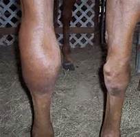
I had the vet run some blood work on Luck and Chance as a precaution, because of the “Panic Grass” in Virginia has been causing liver failure in horses, and because I like to do a full work up every 6-12 months.
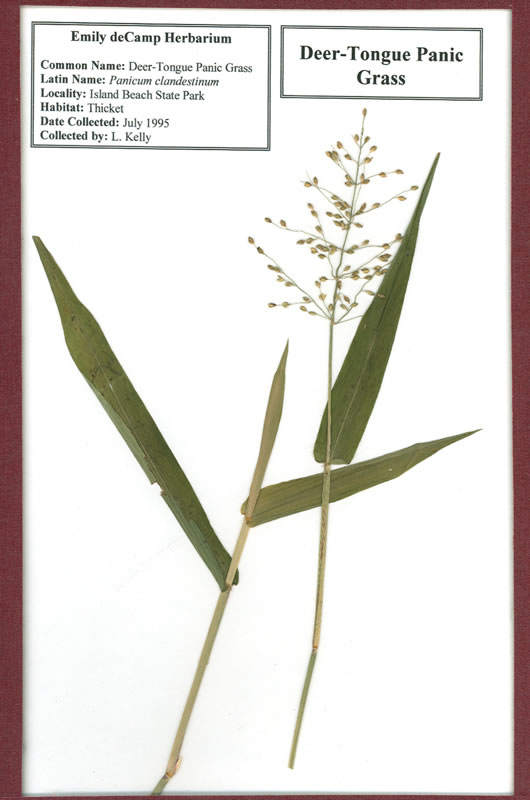


Chance’s Blood Work


C 1

C 3


L 2


INTERPRETATION OF RESULTS


Elevated Potassium (6.6 mEq/L):

“Low levels indicate depletion and are often a predisposing factor, along with
dehydration, in fatigue, muscle cramps, colic, synchronous diaphragmatic flutter (“th
umps”), diarrhea and other symptoms of exhausted horse syndrome. Even seemingly normal or high-normal levels may in reality be lower, but appear higher due to concentration secondary to dehydration as measured by total protein and albumin levels.
Therefore, levels at the lower end of the normal range should be evaluated relative to concurrent dehydration” (https://www.quia.com/files/quia/users/medicinehawk/2407-Vet/labs.pdf).

Low Sodium:

“Low levels commonly indicate loss through excessive sweating, or through kidney or intestinal disease. Low levels may also be found in young foals with bladder damage. Increased sodium levels are usually a sign of dehydration” (http://www.minstervets.co.uk).

Low Platelets: 

This was the most worrisome in regards to the potential immediate issues that could ensue because of the low platelet count. 

“The platelets are the third cellular component of blood (along with red and
white blood cells). These cells contain a number of biologically active molecules that are
critical to the blood clotting process. Low levels may indicate a number of disease
processes not necessarily directly related to a bleeding disorder. Chronic or acute blood
loss, immune disease, toxemia, liver, spleen or bone marrow disease, or even critically
reduced or increased body temperatures can also cause low platelets counts. Any
significantly low platelet counts should be further investigated by a veterinarian. High
levels are generally clinically insignificant unless the condition persists, in which case it may be indicative of bone marrow neoplastic disease” (Susan Garlinghouse).

Low Glucose:

“Glucose is the source of the body’s energy. It is measured in suspected cases of equine metabolic syndrome and sometimes in cases of equine Cushing’s disease. Blood glucose may also be measured as part of a glucose tolerance test, assessing small intestinal function” (http://www.minstervets.co.uk).

Chance was tested for Cushing’s Disease within the last year and the test showed that he did NOT have Cushings. 
Elevated CPK (337 U/L):

Levels 2-3x the highest number in range are considered significant according to vetstream.com.  Levels are easily increased due to poor handling techniques as well as lab error.

According to Dr. Christine Woodford and Carla Baumgartner on vipsvet.com, “Elevations of CPK and SGOT are indictors of muscle inflammation–tying-up or rhabdomyolysis. The term “rhabdo” means muscle and “myolysis” means rupture of muscle cells. The CPK and SGOT are very sensitive indicators of skeletal muscle damage, and they rise in concentration proportionally with the amount of damage. A bit of timing is required in order to obtain the most sensitive results; CPK rises (due to its leakage from muscle cells into the blood system) approximately six to eight hours after the onset of muscle inflammation, and SGOT rises after approximately 12-14 hours. The absolute peak of CPK concentration and the time it takes to return to normal are important indicators of the severity of muscle damage and the response to therapy.”

Elevated MCV: Is the average volume of red blood cells.

  • Macrocytosis.
  • Indicates immature RBC in circulation (suggests regenerative anemia).
  • Very rare in the horse, but may observe increasing MCV within normal range as horses increase erythropoiesis.

According to Vetstream.com, “Macrocytosis (increased MCV) resulting from release of immature RBC from the bone marrow during regeneration is very rare in the horse therefore the MCV is less useful in the horse than in other species.”

Elevated MCH: Is the average amount of hemoglobin in an individual red blood cell.

  • Hemolysis, if intravascular in nature .
  • Errors can occur during processing

Low RBC:

“You may be inclined to think that red blood cell levels need to drop significantly before they cause a problem for your horse. But the truth is that even low-grade anemia – levels hovering around that 30% range on a PCV – can impact your horse physically and may indicate a health problem. This is especially true for high performance athletes. The greater your horse’s physical condition and demand, the higher on the range of normal her red blood cell counts will typically be. Therefore, a red blood cell level low on the normal range or just below may indicate a concern for a racehorse, for example, where it wouldn’t for that pasture pet.”  See more at: http://www.succeed-equine.com/succeed-blog/2014/02/05/anemia-horses-part-1-just-equine-anemia/#sthash.JJuWN5ob.dpuf


Luck’s blood work


L 1

C 2

Elevated Potassium: Potassium can become elevated for a number of reasons.

According to Vetstream.com,

  • 98% of potassium is intracellular.
  • Changes in serum or plasma potassium levels reflect fluid balance, rate of renal excretion and changes in balance between intra- and extracellular fluid.
  • Hypokalemia increases membrane potential, resulting in hyperpolarization with weakness or paralysis.
  • Hyperkalemia decreases membrane potential with resulting hyperexcitability.
Susan Garlinghouse states that, “High serum levels of potassium during an endurance ride are generally not a concern. These increases often reflect nothing more serious than a delay between blood collection (when potassium is actively sequestered inside cells) and sample measurement (after potassium has had time to “leak” from inside the cells out into the plasma or serum).”  This could also be a result of Luck and Chance running around in the heat when the vet arrived.
Increased [potassium] (hyperkalemia) can occur from;
  • Results can be false due to processing time (ie: if the lab waited too long to process blood sample)
  • Immediately after high intensity exercise.
  • In association with clinical signs in horses with hyperkalemic periodic paraysis (HYPP)    .
  • Bladder rupture (neonate)    .
  • Hypoadrenocorticism  [Pituitary: adenoma]  (rare).
  • Metabolic acidosis.
  • Acute renal failure    .
  • Extensive tissue damage (especially muscle).
  • IV potassium salts, eg potassium benzyl penicillin, potassium chloride    .
  • Phacochromocytoma (rare in the horse).
    Hypokalemia
  • Chronic diarrhea.
  • Diuretic therapy, especially potassium-losing diuretics.
  • Excess bicarbonate/lactate therapy.
  • Chronic liver disease    .
  • Acute renal failure (polyuric phase)    .
  • Recovery from severe trauma.
  • Metabolic/respiratory alkalosis.
  • Prolonged anorexia.
  • Recovery period after high intensity exercise (30-60 min after).
  • Parenteral feeding.

In combination with clinical signs and results of other tests results could signify the following;

Elevated GGTP:

* Donkeys tend to have 3x higher levels then horses.  This means that in stead of the typical equine range being 1-35 U/L a typically donkey’s range would be up to ~105 U/L.  Lucky’s test showed he had 120 U/L which is still elevated but not much.  It took sometime to get Luck from the field when the vet arrived- he ran around non stop.  The excitement and anxiety could be the cause of the elevated levels.

RBC:

Katherine Wilson, DVM, DACVIM, of the Virginia–Maryland Regional College of Veterinary Medicine (See more at: http://equusmagazine.com/article/decode-horses-bloodwork-27122#sthash.sc4J1ISJ.dpuf) explains “RBC count is probably the least helpful information because horses usually don’t have big changes in red blood cell numbers. It is not uncommon for horses to have an RBC count a little lower than normal range, however. The term we use for low RBC is anemia, but unless the count gets very low, a horse doesn’t necessarily need to be treated for that condition. A lot of diseases or any chronic long-term disease can cause mild anemia. Usually if we see mild anemia on the bloodwork and the horse has other issues, the anemia is just an indication that we need to fix/treat another problem.”

Low or Elevated Values

  • Splenic contraction.
  • Polycythemia (rare) .
  • Dehydration.
  • Consider causes of anemia
  • Blood loss    .
  • Hemolysis (i in vivo or artifact).
  • Decreased bone marrow production.
  • Poor technique at sampling.
  • Poor handling and storage of samples.
  • Poor technique in laboratory.

Low Bilirubin:

Heather Smith Thomas of Equus states,  “Another indication of liver health is a pigment called bilirubin, which is formed from the breakdown of red blood cells. Elevated levels can mean unusual loss of red cells or liver dysfunction. However, in horses, unlike other animals, elevated levels of bilirubin often isn’t serious. “This value can increase fairly rapidly when horses go off feed, and this is something that is unique to the horse,” says Wilson. “Often we get phone calls from veterinarians who don’t work on horses much or owners who see the blood work and note that the bilirubin is above normal range and are concerned about liver disease. If the horse is off feed for 24 to 48 hours, that value will increase, but this is just a temporary elevation.”

Elevated Hemoglobin (19 g/dL):

According to vetstream.com, Thoroughbred and other “hot-blooded” horses Hemoglobin range differs from other equine- the thoroughbred range = 11.0-19.0g/l.

Elevated Hematocrit (54 %):

 Elevated levels could be due to;

  • Dehydration.
  • Splenic contraction.
  • Polycythemia .

“A measurement of the relative amount of red blood cells present in a blood
sample. After blood is drawn, a small tube is filled and centrifuged to separate the heavier
blood cells from the lighter white blood cells and the even lighter fluid (plasma or serum)
portion. A higher than normal reading generally indicates dehydration (same number of
cells in less plasma volume) or may be due to splenic contraction secondary to
excitement or the demands of exercise. A low reading may indicate anemia, though not
invariably. Highly fit athletic horses may normally have a slightly lower hematocrit at
rest due to an overall more efficient cardiovascular system. Evaluation of true anemia in
horses requires several blood samples over a 24-hour period” (Susan Garlinghouse, 2000/ http://www.equinedoc.com/PrideProjectInfo.html).

It took sometime to get Luck from the field when the vet arrived- he ran around non stop.  The excitement and anxiety could be the cause of the elevated levels.

Low Sodium:

According to horseprerace.com, “Low levels indicate depletion and are often a predisposing factor, along with dehydration, in fatigue, muscle cramps, colic, synchronous diaphragmatic flutter (“thumps”), diarrhea and other symptoms of exhausted horse syndrome. Even seemingly normal or high-normal levels may in reality be lower, but appear higher due to concentration secondary to dehydration as measured by total protein and albumin levels. Therefore, levels at the lower end of the normal range should be evaluated relative to concurrent dehydration.”


More information on your horse’s blood work

Decoding your horse’s blood work

CBC and Chemistry Profile

A Better Understanding of the Results


The vet suggested that I add water to Luck’s and C’s feed in case their values are due to dehydration. She also explained that some of the values may be a result of running around in the field right before drawing them along with anxiety.  

The anxiety and running around seemed fair but I am hesitant on the dehydration portion.  Yes, I know it is winter and that horses are less likely to drink as much water.  But if it were due to dehydration then the Albumin would be low as well.  But, the blood work revealed that the Albumin was 2.8 (Luck) and 3.2 (Chance).  These values are within the normal range…. that being said, the results could also be due to lab handling especially the Potassium levels.

While speaking with my uncle Jerry (the horse whisperer), he suggested adding a salt block to the horse’s feed.  This will increase the horse’s thirst which will get them  drinking more.  I also added heated water buckets so that the water won’t freeze and in case they are less inclined to drink when the water is cold.

In order to feel comfortable about my horse and donkey being healthy, I will have more blood work done this week to make sure everything is in fact okay.


Laser Time

A little over a year ago Chance was diagnosed and treated for a lesion on the Deep Digital Flexor Tendon (DDFT) of his hind fetlock.  At the time, I decided to go with Stem Cell injections due to it being the least invasive treatment option.  The Stem  Cells worked wonders and Chance went from barely being able to walk to running up hills, from daily pain medications to a supplement (DuraLactin) to help with pain and inflammation) for arthritis.

A few months ago I wanted to see how Chance’s fetlock was holding up.  He did not have any symptoms- no swelling, lameness, etc- but I wanted to make sure.  The ultrasound revealed that he did still have a small lesion on his DDFT that the vet felt was “congenital” and not problematic.  But due to his history I wanted to ensure that it stayed non-problematic.  I invested in some protective and supportive hind boots for hi to wear when he is outside or stall bound due to weather and decided to do a round (about 5 sessions) of laser treatments on his hind leg.  The laser treatments are relatively inexpensive (about $65.00), non-invasive, and pretty much risk free.

We are coming up on our 3rd treatment and due to Chance being asymptomatic it is hard to tell if they are beneficial until we do another ultrasound.  Once an ultrasound is completed I will post more.

Article on Stem Cell Treatments:  http://www.rossdales.com/news/new-stem-cell-trial-to-treat-deep-digital-flexor-tendon-injury.htm

Articles on Laser Treatments: Research Laser Therapy


Horses in Virginia are at risk of Liver failure due to Panicgrass or Panicum.

Horses in Virginia are at risk of Liver failure due to Panicgrass or Panicum.

IMG_1763-1030x773

According to Haymarket Vet (http://haymarketvet.com/fall-panicum-grass-and-liver-disease/) Panicgrass is causing Fall Panicum Toxicosis in horses.

“In 2004, our practice was involved in documenting an important toxin for horses—fall Panicum (Panicum dichotomiflorum) grass.  This common native grass has been fed to horses in hay and in pasture probably since the Europeans first brought horses to our area.  But, while we know that it doesn’t cause illness all the time, certain growing conditions can cause it to become toxic, as it did in Nokesville, VA in 2004.  We don’t know what triggers the grass to become toxic, but we do know that it sometimes does become toxic, and the conditions are right this year. This study proved the hepatotoxicity: http://www.ncbi.nlm.nih.gov/m/pubmed/17186859/

Currently, there are several cases of liver disease in Fauquier, Clark and Loudon counties that appear to be from grazing Panicum grass in the pasture. Some signs of toxicity from eating the grasses includes: decreased appetite, lethargy, somnolence (unusual periods of sleepiness), mild colic, or neurological signs. Some horses have no symptoms at all.

If you have this plant in your pasture or if you find it in your hay cut this year, you may want to have your horses tested for liver disease; this involves a simple blood draw.”

Below is a link on more information about Liver Disease, symptoms, and treatment options.

http://www.tsln.com/agliving/equine/7614069-111/liver-horse-says-disease

Floating Away

Lately, I have noticed that Chance is eating more on the left side of his mouth and is taking a longer time finishing his meal.  I booked an appointment with the dentist and he came out to take a look.

Chance showed discomfort while being floated on the right side of his mouth.  However, upon taking a closer look the dentist did not see or feel any loose teeth nor did he smell an odors (indicating a broken or infected tooth).  I asked if Chance could have a tooth that was cracked near the root?  The dentist explained that this could be the case and we would need to do an x-ray to know more.  He suggested that we see how he does after being floated, due to one of his teeth on the upper right side being higher than the others, and go from there.

The dentist did say that Chance’s teeth were sturdy and in good shape especially given his age! Yay!